| OCR Text |
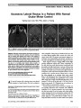
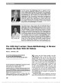
Show Intermittent Horner Syndrome in a Pediatric Patient Meenakashi Gupta, MD, Ilya Leskov, MD, PhD, Joshua M. Kruger, MD, PhD, Dean M. Cestari, MD Abstract: Intermittent Horner syndrome is uncommon in both the adult and pediatric population. We describe a case of a pediatric patient with an intermittent Horner syndrome. Infrared photography and videography were used to help establish the diagnosis. Journal of Neuro-Ophthalmology 2014;34:149-150 doi: 10.1097/WNO.0000000000000062 © 2013 by North American Neuro-Ophthalmology Society A14-year-old healthy boy was evaluated for a history of episodic right upper eyelid ptosis accompanied by mio-sis of the right pupil. His parents noted this on 4 occasions in the week preceding his office visit. Each episode lasted 15-20 minutes and resolved completely. There was no associated headache, rhinorrhea, tearing, nasal congestion or anhidrosis, nor a history of head or neck injury. Two weeks before presentation, the patient was struck in the right eye with a snowball. At that time, he did not seek medical attention. His mother reported some mild swelling of the right eyelids that resolved within 1 week. Initial neuro-ophthalmic testing was unremarkable with-out ptosis or anisocoria. During the course of the examina-tion, the patient developed a mild right upper lid ptosis and miosis of the right pupil (Fig. 1A). Infrared videography revealed right pupillary dilation lag (See Supplemental Dig-ital Content 1, Video 1, http://links.lww.com/WNO/A88). The ptosis and miosis resolved after 20 minutes (Fig. 1B), as did the pupillary dilation lag (See Supplemental Digital Content 2, Video 2, http://links.lww.com/WNO/A89). The ocular and neurological examinations were otherwise normal, and no triggering maneuvers were identified. Magnetic resonance imaging (MRI) and magnetic reso-nance angiography of the brain were unremarkable. MRI of the neck revealed a 1.7-cm cervical syrinx extending from C5 through C7, which spared the interomediolateral cell columns (Fig. 2A). MRI of the neck also revealed 2 nodules in the right portion of the thyroid gland. Thyroid ultrasound demonstrated 2 heterogeneous solid nodules, one measuring 0.8 cm · 0.9 cm · 2.0 cm in the right upper pole and the other measuring 0.6 cm · 0.8 cm · 0.9 cm in the right midpole. Neither nodule was in contact with the sympathetic chain. Thyroid function studies were normal, and ultra-sound- guided needle biopsy of the thyroid nodules revealed FIG. 1. A. There is right upper lid ptosis and right pupillary miosis. B. The lid and pupillary findings have resolved. Department of Ophthalmology (MG, IL, DMC), Massachusetts Eye and Ear Infirmary, Boston, Massachusetts; Neuro-ophthalmology Service (DMC), Department of Ophthalmology, Massachusetts Eye and Ear Infirmary, Boston, Massachusetts; and Department of Ophthalmology (MG, IL, DMC), Harvard Medical School, Boston, Massachusetts. Supported by the Medical Scientist Training Program, Grant T32GM007753 (IL), from the National Institute of General Medical Sciences. The authors report no conflicts of interest. Supplemental digital content is available for this article. Direct URL citations appear in the printed text and are provided in the full text and PDF versions of this article on the journal's Web site (www. jneuro-ophthalmology.com). Address correspondence to Dean M. Cestari, MD, Neuro-ophthalmology Service, Department of Ophthalmology, Massachusetts Eye and Ear Infirmary, 243 Charles Street, Boston, MA 02114 Gupta et al: J Neuro-Ophthalmol 2014; 34: 149-150 149 Photo Essay Section Editor: Timothy J. McCulley, MD Copyright © North American Neuro-Ophthalmology Society. Unauthorized reproduction of this article is prohibited. no signs of malignancy. Three months after initial presenta-tion, the patient reported less frequent episodes of ptosis and miosis that totally resolved 6 months after initial presentation. Our patient's findings are consistent with an intermit-tent Horner syndrome. The fluctuating clinical course is presumably caused by a reversible functional impairment of the oculosympathetic pathway to the right eye. Although intermittent Horner syndrome has been associated with cluster headaches and spinal cord lesions (1-4), the etiology in our patient is uncertain. The presentation of Horner syndrome related to cluster headache is variable and, in some cases, its onset precedes the development of headache by several years (5,6). Other cases have been described in which patients experience no pain yet develop miosis, ptosis, and other accompanying symptoms without experiencing pain including rhinorrhea, nasal stuffiness, and lacrimation. This entity is known as cluster headache sine headache (6-8). Episodic Horner syn-drome in the absence of headache, rhinorrhea, nasal stuff-iness, and lacrimation also has been reported (3). This case was suspected to be a cluster headache variant (3). Another consideration for the intermittent Horner syndrome observed in our case is the patient's history of ipsilateral ocular trauma. The Horner syndrome began 2 weeks after the trauma and resolved after 6 months. However, the ocular injury could have theoretically resulted in neck and vascular damage that led to the development of the ipsilateral intermittent Horner syndrome. However, no structural damage was detected by neuroimaging studies. Thyroid pathology has been associated with Horner syndrome when the cervical sympathetic pathway is affected (9). However, ultrasound revealed that the thyroid nodules in our patient did not compress the sympathetic chain. Syrinx of the cervical spinal cord may be a cause of Horner syndrome. This is because of dysfunction of the second-order neurons in the interomediolateral cell column in the C8 to T2 levels (10,11). Intermittent dysfunction of the oculosympathetic pathway has been reported from an acquired syrinx (12). The syrinx in our patient appeared centrally located in the cervical cord. Yet, it had a similar appearance to the syrinx reported by Pomeranz (11), in a patient who developed a right Horner syndrome. Fluctuations in the size of the syrinx or shifting fluid within it may have caused the intermittent Horner syndrome in our patient. REFERENCES 1. Hopf HC. Intermittent Horner's syndrome on alternate sides: a hint for locating spinal lesions. J Neurol. 1980;224:155-157. 2. Zur PH. Intermittent Horner's syndrome: recurrent, alternate Horner's syndrome in cervical cord injury. Ann Ophthalmol. 1975;7:955-962. 3. Murphy MA, Hou LC. Recurrent isolated Horner syndrome. J Neuroophthalmol. 2006;26:296. 4. Pollard ZF, Greenberg MF, Bordenca M, Lange J. Atypical acquired pediatric Horner syndrome. Arch Ophthalmol. 2010;128:937-940. 5. Havelius UA. Horner-like syndrome and cluster headache. What comes first? Acta Ophthalmol Scand. 2001;79:374-375. 6. Salvesen R. Cluster headache sine headache: case report. Neurology. 2000;55:451. 7. Dilli E, Dodick DW. Extracephalic cluster (cluster sine headache). Neurology. 2008;70:1362-1363. 8. Leone M, Rigamonti A, Bussone G. Cluster headache sine headache: two new cases in one family. Cephalalgia. 2002;22:12-14. 9. Harding JL, Sywak MS, Sidhu S, Delbridge LW. Horner's syndrome in association with thyroid and parathyroid disease. ANZ J Surg. 2004;74:442-445. 10. Kerrison JB, Biousse V, Newman NJ. Isolated Horner's syndrome and syringomyelia. J Neurol Neurosurg Psychiatry. 2000;69:131-132. 11. Pomeranz H. Isolated Horner syndrome and syrinx of the cervical spinal cord. Am J Ophthalmol. 2002;133:702-704. 12. Kline LB, McCluer SM, Bonikowski FP. Oculosympathetic spasm with cervical spinal cord injury. Arch Neurol. 1984;41:61-64. FIG. 2. T2 axial (A) and sagittal (B) magnetic resonance imaging show a syrinx (arrows) at the C7 level. 150 Gupta et al: J Neuro-Ophthalmol 2014; 34: 149-150 Photo Essay Copyright © North American Neuro-Ophthalmology Society. Unauthorized reproduction of this article is prohibited. |