| OCR Text |
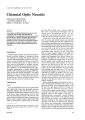
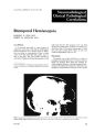
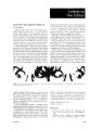
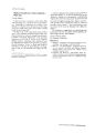
Show f. Clin. NeuflHlphrh.llnIl1/. 2: 133-13<l. I <l1l2. Neuroradiological Clinical Pathological Correlations Bitemporal Hemianopsia ROSENDO D. DIAZ, M.D. ROBERT M. QUENCER. M.D. Case History A 17-yeolr-l)ld molle with iI l-ye.u history of progressive visil1n loss in the left eye and frequent l>ifrontoll he.ldolches for () months prior to admission W.lS found on examination to have a visual acuity on the left of 20/100; optic pallor WilS evident on the right. The visual fields revealed an asymmetric bitemporal hemianopsia greater on the f r"m tht' Dt'pMlmenl "f R..diology. Neuwr..diology Div, ,('n. L'n"'ers'tv of Ml.. m'.I.. cl..son Memorial Mt'dlCdl Center. M,..m•. FI"nd. right than the left. The prolilctin level was 7810 ng/ml. The radiographic evaluation included plain skull films (Fig. 1), computed tomography in both the axial and coronal plains (Figs. 2a -2d), and cerebral angiography (Figs. 3a -3c). Discussion The lateral view of the skull (Fig. I) reveals an enlarged and eroded sella turcica. Common entities which can produce these plain film findings include: pituitary adenoma, craniopharyngioma. metastasis to the sella and/or pituitary, sphenoid sinus neoplasm, intracavernous aneurysm of the carotid Figure I. wleroll view of the skull rev<'db b.. I1"'>nin}; "f th,' ",II" lurek" wilh en"i,,,, "f Ihe ,,'II,1r 11",'r. th,' "nJN<urf,KC' of b,'lh anlerior clinoids. olnd the dorsum ,ell. (drww,j. N" -urr.. or i"lr"",II... , .,1, if"'"li"" i, ,J"l1lifil'd "II pl,lI" ,I..ull film,. June 1982 133 Figures 24-2(, Prl"lllntr,ht (T "(..111 t.11 ..h'IW" .1 1"bul.lt~J "'urr~l:-dl.,r m.I~~ ''\'lth JfC'..h ,... t .....Jlohl·.Jtl\ln ... 1"'lrJ.,).:ht .lrrl'\\'~1 ..lnd 1\ w .111l'nU.'lI,'" IcurV(·J Mn'wl. P,'<IC"nlrJ-t ~<,.In ," Ih" ."'J! (I:>. ,'1 ,'nd c,'rl'nJI rIJnl'- IJI. ,h,.\\- tIll' prE'-"n,,, d Ih" mJ-- In th(' ,,('IIJ (\I .",J ,,~ht rJrJCJVE'rn,'u~ r"~I('" (,.p"" Jrn'wl wllh S1bnifoc.,nt r,><I"",'r ('\I"n"Il'n I" Ih" kn'I"t b"th ("r"brJI p"duncl".; lyJ Th(' "'r,'n,11 CJn I d, dr,lmJlI(Jllv d('m,",.lrJI"~ Ih" "urNIl'r l'\t"nt "f lh(' m.l>" In \\ hl<'h Ih"r" hJ" b('"n br,,,\·th ,nt" th" nght r flvrnlru.. ul ..H r(,~l\'n (white' ..1rn'\V~) \It(' In thC' P\'~I"""ntr.hl "'l.ln thC' Inh\.'m,'~('n{'\.'u~ cnh.ln,'cm('nt JnJ the cystl~ l·l"'mp,'oenls. Jrtery. dnd suprJSelldr meningiomol. Other dise.1se processes such dS an optic chiolsm or hypothalolmic gliomol. germinom,l. hypotholl.:lmic histiocytosis. enlargement of the anteril'r portion l,f the thi rd v('ntride. holmartomJ.•md primolry hvpl)thyr,)idIsm in children would only r,Hely result in this Jpp('.H.ln e. Supr.1s('lIolr meningiom.1s r.uelv l)CCUr olt thi. agel Jnd hypero. tosis.•1 common fe.1ture l,f <'uprJ~('IIJr m('ningil)m.1s det('cted in 50",{, l,f C.l. ('s.! i~ nut pre'. ('nl. An intr.1CJVernllUs .ulC'llrysm l)f th(' intNn,l1 (.Iwtid .Irt('ry in .1 p.1tie'nt l,f this olgr whl' h.ld no hi.,tory uf tr.1Um.1 would b(' unu. U.ll.·' Furthl'rmure. introlcrolni.ll.lneurysms in p.ltients <'f this Jge' t('nd to be found periphN.1I1y. di.t.11 tt) the (irclt' llf Willis. In addition. on pl.lin films .m intrac.lVrrnou<, an('urysm produ('s .1n .1symnH'tric. ll l'nlolrgrml'nt of thr srll.l with molrkrd ('wsil)n of lllll' .lntC'rillr dinoid. An l'nl.Hged .1l1trrior pl)rti( lll (If the third vrntriclr will rnl.trge the 5('11,1 but will .11 .." lkmilH'r.lli7l' .lnd N"d(' thr postrrior di"" rt!· .lntl till' "'!' ..t th\· J"r<,um, .1 finding not present in this patient. Primary hypothyroidism in children. prl,bJbly due to overactivity of the pituitJf\' gl.1nd. I will enlarge the sella but not to the degree seen in this CJse Jnd will not cause bone destructilm. The CT SCJn (Figs 2J -2d) shows an intrasellar .lnd supr.1sellJr 10buIJted m.lSS with rim calcificatil'ns ,1l1d inhomogeneous enhancement. On the b.1Sis of these CT findings we can definitely exclude .1 nl'nthrombotic or partially thrombotic .1l1eurysm.•md In enlarged anterior portion of the third ventricle. A supr.lsellar meningioma is very unlikelv since hyperostosis. which is seen on CT in N"""~ of supr~sellar meningiomas is lacking'; there is .m inhomogeneous enhancement rather th.1n the typical sharply defined homogeneous enholncement of meningiomas. and there is extensive bone destruction. which would be unusual for a meningioma. Although an optic chiasm glioma. which is predominantly a tumor of early childhood, mJy have similar enhancing char.lcteristics as in Journal of Clinical Neuro-ophthalmology June 1982 (c) 135 Figure 1 (e,lOtlflUcd, (d) I'iKure, -'a--'c. "II''''''I11I''I'''d AI' ,'1 till' nj;hl .",.1 1..11 '''','lid ,1<I,·rl<'j;r.1I1" (.11. presenhng evidence of l.ller"l displacement of the I',,,"'n,.r '''1',',1 "I till' nght ''''tlt,d "I'h"" (Mr,'wl \\'h,d, " Ih(' H'V('r-,' "i 'wrn",1 "nd indicates laleral e"tension of the inlrasellar ,,,",p,,m'''1 "I th,' ,".," H,.th ".1 '1'j;I\"'I\I' ,., th .. ""I,'r\\'r lI'H'br,11 "'Icries .lr(' displaced superiorly (open arrows) indicating the ",.",', .,,,I",,,,r .11,,1 '1'p,'r".r 1"1"",,,.,,. Arla,.,1 ph.",· <I" Ih,' I"tcr.,I VIew (h-,'nly on(' ide shown) shows a forward di placement ,,' Ih,' '''I'r." 10"",,1 I'"r""" "I Ih,' ",la".,1 ,.lr,'I,d "'lay (bl,,,"I.. .Irr,'wl. ""pen",g Ihe siphon." Tumor arteries (white arrow) 1111):IU.IIlIL): 1"lnl lhf" I ,1\1"II\I1U" l.1fl'tut .Ht" It'.HI\' ,,,'('11 .\nd the' rt":-.ult uf thiS ..lbnormal v.Jscularity is a faint tumor stain (surrounded 1.\ '01" III HI •• \,\ 'iii Ill" \1 '1"11'. ph., ... I. 1,,1 Ihl' ,\Il).:,hlgr.\l1l Journal of Clinical euro-ophthalmology , , .< ("'J our case, they do not contdin the> rim c,llcific.Jtion!'> or cystic components as in Figure 2, A hypothJIJ. mic glioma presents as d suprasellar mdSS, hypodense, isodense or hyperdense tu urrounding brain, but they usudlly contrdst enh.1I1c(' to J greater degree thdn in our patient," .lnd (on!'oidl'r- June 1982 .1ble- vl'ntriclIl.H Jil.lt.lti'"1 dill' h) in\'.l~i,)n "f the for.lllll'n llf Mllnn) WllUld bl' St'l'n in .1 hvpothJI.1mil' gliom.l of thi. SiZ('.7 A gl'rmil1l)mJ J11.{y .Hise in the' rl'gion of the- hYPl)th.ll.1J11US but f.Hely do they inv.lde- the pituit.lry gl.lnJ to produce the rddiogr. 1phil- finding. s('('n in l)Ur p.1tienl. Hypothdlamic 137 Bit(·mplIr.11 HC'mi.1I111psi.1 histiocytosis presents ,1S .1 large suprasellar mass, with hypodense areas in the hypothalamus, and m.lY contr.lst <>nh.mce" but do not contain calcifi(:' ltl(1I15. Furthcrmor(', both germinomas and hypllth. 1l.tmic histiocytosis frequ('ntly C,lUSP diabetes insipiJus. Hypoth.ll.lmic ham,lrtomas unusually ewd(' the 5('11.1, .llthough the CT .lppearance of the m.1SS itself m.1Y be identical to our case; however, th('s(' h.lm.Htl1m.lS r.lrely exceed 2 cm in diameter .1I1d thl' p.llients ,llmost invariably present with precocious puberty.' Neither a metastasis to the sphenoid .lnd pituitary or a primary sphenoid sinus neoplasm would show calcification on CT and in addition, by the time a sinus carcinoma became large enough to cause such an intracranial mass, extension into the adjacent sinuses, nasal cavity, or nasopharynx would have been evident. The two most likely diagnoses, then, would be craniopharyngioma and pituitary adenoma. Characteristically the CT findings in childhood craniopharyngiomds are a suprdsellar mdSS which in some cases may have an intrasellar extension. Calcifications are seen on CT in gredter than 80% of the cases:1 Typically the calcifications are rim-like which mdY sometimes extend beneath the diaphragma sella. Contrast enhancement (75% of cases) and cystic components (50% of cases) are more common in children," than in adults. When the cystic component enhances it is usually the cyst wall which enhances. The cystic areas may encompass the entire tumor or a small volume of it. Fitz" found that a centrally placed suprasellar tumor in children with two or more of the following three CT findings-contrast enhancement, tumor calcification, or cyst formation-was a craniopharyngioma in eight out of the 10 cases. Our patient has all three findings, so that a craniopharyngioma based on CT would be a good diagnostic possibility. The typical CT findings in pituitary adenomas are an intrasellar mdSS, with or without suprasellar extension, producing enlargement of the sella with equal or slightly higher attenuation than the surrounding brain. The tumor enhances, usually homogeneously, in 55% of the cdses,r. dnd may possess lobulated or irregular borders. Atypicdl appearances include cystic degeneration (7%).~· invdsion of the anterior, middle or posterior cr.midl foss.l, dnd tumor calcification (3%-7%).\' Therefore, the CT findings in our patient may represent an atypically appearing pituitary adenoma. The final radiographic study performed WJS .1 cerebral angiogram with nlclgnific.1tion <lnd subtraction (Figs. 3.1 -3c). It showed .I supr.lsellilr extr.1axial mass extending into the subfront.11 region. In addition, two diagnostic findings Jlmost never seen in craniopharyngiomJs but found in pitUitclry ad- 111.11 • enomas were s('en. One IS the presence of tumor .:ITtNil's originating from the .mterior capsule artery and the second is a tumor stain. Thus, based on the CT and angiographic findings, a pituitary adenoma was felt to be the most likely diagnosis. A right frontotemporal craniotomy was perfonned for subtotdl removal of the tumor. The final pathological report was aggressive pituitary adenoma. Jefferson I~ was the first to report on invasive pituitary adenomas. These pituitary adenomas extend beyond the pituitary capsule, infiltrating and destroying adjacent structures, but as opposed to pituitary carcinomas, they do not metastasize. Lundberg':' reviewed 11 patients with aggressive pituitary adenomds and all had very high serum prolactin vdlues, a finding observed by other investigdtors.'~ It is speculated that the very high serum prolactin level is caused either by autonomous hormone secretion from the large number of abnormal cells or is secondary to invasion of the cavernous sinus by the prolactin-secreting tumor.I~>. IH References I. Richmond, I.L.. and Wilson, C.B.: Parasellar tumors in children. I. Clinical presentation, preoperative assessment. and differential diagnosis. Child's Brain 7: 73-84, 1980. 2. Di Chiro, G., and Lindgren, E.: Bone changes in cases of suprasellar meningioma. Actd. Rddio/. 38: 133138. 1952. 3. Hammock, M.K., and Milhorat, T.H.: Cranial Computed Tomography in Infancy and Childhood, Williams and Wilkins, Baltimore, 1981. 4. Van Wyk, J.J., and Grumbach. M.M.: Svndrome of precocious menstruation and galactorrhea in iuvenill:' hypothyroidism: an example of hormonal overlap in pituitary feedback. /. Pediatr. 57: 416, 1960. 5. Banna. M., et al.: Pituitary and parapituitary tumors on computed tomography. A review article based on 230 cases. Br. ]. R.!diol. 53: 1123-1143, 1980. 6. Miller, J.H., et al.: Radiological investigation of sellar region masses in children. Radiology 134: 81-87, 1980. 7. Lin, S.. et al.: Radiologic findings of hamartomas of the tuber cinereum and hypothalamus. Rddio/ogy 127: 60 7-703. 1078. 8. Fitz. C.R.. et al.: Computed tomography in craniopharyngiomas. Radiology 127: 687-691, 1978. o. Lmdolt, A.M., and Rothenbuhler, V.: Pituitary adenom. l c.1Icification. Arch. Patho/. Lab. Med. 101: 2227, 1077. 10. B.1ker. H.L.. Jr.: The angiographic delineation of sellar dnd paraselldr mdsses. Radiology 104: 67-78, 1072. II. Numaguchi, Y., et al.: Neuroradiological manifestalions of suprasellar pituitary adenomas, meningiomas. and craniopharyngiomas. Neuroradiology 21: 07-74, 1981. 12. Jefferson. G.: Extrasellar extensions of pituitary adenomas. President's address. Proc. R. Soc. Med. 33: 433-458, 1940. 13. Lundber, P.O., et al.: The invasive pituitary ade- Journal of Clinical Neuro-ophthalmology noma. A prol.1l'tin producin~ tumM. An-h. Nl'url,/. 34: 742-740 , 1°77. 14. Malarkey. W.B...md lohnslll\. I.e.: I'ituil.,ry lun1<lr~ and hypprprol.Ktil\('mi.1. Arch. Intl'rn. Met!. 13t>: 4044, 10 70. 15. Shucart, W.A.: lmpli(.1Iil1I\s llf vl'ry hi~h ~t'rUIl1 I'rt'· laclin levels .lsslKi.llE'd with piluil.1ry lun1tlr~. I. Neurosurg. 52: 22(1-228, 101\0. June 1982 Di.lZ, Quencer 1(1. V.1~silllulhi." I., .md Richardson, A.E.: Prolactin l('v('ls in .1~gr('s iv(' piluildry lumors. ]. Neurtlsurg. 53: 131- 132, I Qt\O. Writ(' (or r('pril1/~ I,,: Rob('rt M. Quenler. M.D., I.kp.nlmC'nl of ({.ldiology, N('urorddiology Division, UnivNsity llf Mi.lmi chool of MediCIne, P.O. Box OloQoO, Mi.1mi. F1ori,b 33101. 139 |