| OCR Text |
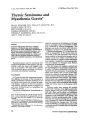
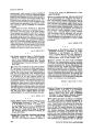
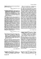
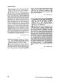
Show ,. Clin. Neuro-oplrt/ramol. 5:254-257, 1985 Thymic Seminoma and Myasthenia Gravis* ISLA M. WILLIAMS, M.D., F.RAC.P., M.RC.P.E., D.O. DAVID C. GEE, F.RC.P.A ERIC COOPER, F.R.A.C.S. PAMELA DICKINSON, M.B.B.S. THOMAS F. SANDEMAN, M.D., F.RAC.R ALEX C. SUM, M. Eng. Sc. <01985 Raven Press, New York Abstract Computed tomography detected an enlarged thymus in a 27-year-old man with myasthenia gravis of recent onset. Pathological examination of the thymus revealed lymphoid hyperplasia and a thymic seminoma (or germinoma), which was arising as an intramural nodule in a thymic cyst. This is the first reported association of thymic seminoma, lymphoid hyperplasia, and myasthenia gravis. Myasthenia gravis may be associated with lymphoid hyperplasia of the thymus, with thymomas, 1,2 and very rarely with Hodgkin's disease of the thymus.3 We report here a patient with myasthenia gravis associated with thymic seminoma and lymphoid hyperplasia. Case Report A 27-year-old man had experienced intermittent diplopia on left gaze in the evening for 6 weeks. Examination revealed weakness of the left lateral rectus muscle on extreme left gaze, with fatigue, weakness of the left orbicularis oculi muscle, and gaze paretic nystagmus to each side. Neither ptosis nor myasthenic lid twitch was present. Intravenous edrophonium hydrochloride re- From the Royal Victorian Eye and Ear Hospital (I.M.W., P.O., A.C.S.), Prince Henry's Hospital (I.M.W., D.C.G., E.C.), and Peter MacCallum Hospital (T.F.S.), Melboume, Australia. • Presented at the Frank Walsh Society Meeting. New Orleans. LA. U.S.A., February 16. 1983, and at the Australian Association of Neurologists Annual Scientific Meeting, Perth, Australia, May 4, 1983. Write for reprints to: I.M. Williams, M. D., 15 Collins Street, Melbourne, Victoria 3000, Australia. 254 suIted in resolution of the diplopia on left gaze and an increased range of movement of both eyes, confinned by infrared oculography. The diplopia was easily controlled with oral pyridostigrnine, 10 mg at 2 P.M. and 20 mg at 7 P.M. Computed tomography (0) of the thorax detected an enlargement of the thymus that was not visible on the plain chest film. The levels of serum antibody to acetylcholine receptor and to striated muscle were within normal limits. The muscle response to repetitive nerve stimulation was negative. Single-fibre electromyography was not available. An enlarged cystic thymus was removed at thoracotomy. After the surgery the diplopia resolved and the pyridostigmine was gradually stopped over 2 weeks. No further abnonnality was found in repeated clinical examinations. In particular, the testes were normal. The cr scan of the body showed no evidence of metastatic disease. The serum levels of a-fetoprotein and of human chorionic gonadotropin were normal. The postoperative course was complicated by left basal atelectasis but was otherwise uneventful. The patient was treated with mediastinal radiotherapy 3,000 rads tumor dose in 20 fractions over 4 weeks. Thirteen weeks after surgery the patient awoke with weakness of the right lateral rectus muscle, fatigue, and gaze paretic nystagmus to each side. There was no ptosis, no myasthenic lid twitch even after prolonged upgaze, and no weakness of the orbicularis oculi muscle. The diplopia resolved spontaneously after 11 days and has not recurred during the 15 months since. Repeated CT scans of the thorax have revealed postoperative changes only. The serum levels of antibody to acetylcholine receptors and to striated muscle 12 months after the operation were normal. Journal of Clinical Neuro-ophthalmology Figurt 1. A low-power \'iew of the tumor nodule and overlying thymic cyst. Note the prominent lymphoid follicles with enlarged germinal centres (GC) and the sheets of tumor ceUs en in the interfollicular zones. CL is the cyst lumen. (Haematoxylin and eosin stain. x 10.) Pathological Findings The operative specimen consisted of a multiloculated thymic cyst, into the lumen of which protruded a well-circumscribed, firm, nodular mass 5 cm in diameter, with a yellow, lobular, cut surface. HistologicaUy this mass was composed of lymphoid tissue showing prominent follicular hyperplasia with numerous germinal Williams et al. centres (Fig. 1). In the interfollicular (sinusoidal) zones were sheets of tumor cells with large irregular oval nuclei having prominent nuclear membranes and traversed by prominent chromatin strands. A single large eosinophilic nucleolus was generally present. The cytoplasm was pale-staining or clear and the cytoplasmic borders ill-defined. There was considerable nuclear pleomorphism, and mitotic figures were plentiful. Small foci of tumour necrosis were present. In many areas the tumor cells were somewhat obscured by the dense lymphoid infiltrate, and aggregates of epithelioid histiocytes indicative of granulomatous stromal reaction were present (Fig. 2). The features were characteristic of a thymic seminoma (germinoma). The associated thymic cyst was lined by focally keratinised, stratified squamous epithelium and contained lymphoid tissue (including germinal centres) and Hassall's corpuscles in its wall. The epithelium partially covered the intraluminal surface of the tumor nodule. Thymic tissue distant from the cyst and tumor also showed lymphoid hyperplasia. Electron microscopy demonstrated ultrastructural features characteristic of seminoma. The tumor cells had a prominent central nucleolus, often with a reticular nucleolonema. The cytoplasm contained few organelles, with the exception of numerous free ribosomes. Aggregates of glycogen were common (Fig. 3). Figurt 1. Higher power of seminoma cells (5) and associated aggregates of epithelioid histocytes and giant cells (GC). Note the nuclear pleomorphism, the large nucleolus and clear cytoplasm of the seminoma cells and areas of necrosis (N). (Hal" matoxylin and eosin, x200.) December 1985 255 Thymic Seminllmo and MyClslhenia Gravis - - Figure 3. Electron micrograph illustrating the seminoma cells with their prominent nucleolus and ~eticula~ nudeol~ne~ (N), sparse cytoplasmic organelles, and aggregates of intracytoplasmic glycogen (G). Bycontrast there 15 a~ adJacent epithelial thymocyte (ET) with numerous cytoplasmic tonofilaments (single arrow) and a pronunent external lamina (double arrow). A lymphocyte (LC) is also present ( x 3.600). Discussion The recent onset of myasthenia gravis may be the first indication of thymic pathology.4 CT following plain chest films has been advocated to differentiate thymic (lymphoid) hyperplasia from thymoma,S especially in the older age group.6 However, CT may yield false-positive results. 7 In the case presented herein CT (but not chest film) identified the abnormally large thymus. Pathological differentiation of thymic (lymphoid) hyperplasia from thymic neoplasia and separation of the various th(miC neoplasms are vital in planning therapy. In contrast to the more common epithelial thymomas, thymic seminomas (like their gonadal counterparts) respond readily to radiotherapy, and the prognosis is excellent.8•9. In view of the clinical presentation with mild ocular myasthenia, it is intriguing that the tumor nodule in our case was composed of seminoma that appeared to have arisen in an area of thymic lymphoid hyperplasia. In our case the lymphoid hyperplasia was present in the uninvolved thymus and cyst wall as well as being intimately associated with the tumor nodule. The most consistent association of thymic pa- 256 thology with myasthenia gravis is lymphoid hyperplasia, whether a thymoma is present or not. I We are unaware of symptoms of myasthenia KTavis having previously been reported as the presenting symptom of thymic seminoma. No such association was described by Polansky et aL, 10 who reported four cases of anterior mediastinal seminoma and reviewed 103 cases reported in the English literature up to 1979. Thirty percent were asymptomatic and detected in routine chest films. In 70% the initial symptoms and signs were characteristic of an invading or expanding mediastinal mass. One can only speculate as to the pathogenesis of myasthenia gravis in this case. The seminoma could possibly be an unrelated incidental finding, but this is unlikely, as the most pronounced lymphOId hyperplaSIa was lnhmately associated with the tumor. There are rare reports in the literature of other neoplasms such as Hodgkin's disease2 and non-Hodgkin's lymphomall being associated with myasthenia gravis. In the latter case, the myasthenia developed follOWing total nodal irradiation of the lymphoma and was associated with a T-cell immunodeficiency. There is no clear evidence that other extrathyrnic malignancies occur with in- Journal of Clinical Neuro-ophthabnology creased frequency either concurrently or after thymectomy for myasthenia gravis,12 but reports suggesting such a relationship do raise the possibility that some underlying thymic abnormality is associated with myasthenia and with the development of systemic neoplasia. The reason for the recurrence of myasthenia gravis symptoms 13 weeks after thymectomy in our patient, followed by spontaneous resolution after a further 11 days, is uncertain. Patients in remission or with purely ocular myasthenia gravis have a lower incidence of elevation of serum acetylcholine receptor antibody titers, whereas patients with generalized severe myasthenia gravis, particularly in the presence of thymomas, tend to have the highest levels. l3 The minimal ocular symptoms in the patient presented are in accord with the normal levels of antibody to acetylcholine receptor and to skeletal muscle. Acknowledgment Support in the form of grants was given by the Australian Brain Foundation, Eye Ear Nose &Throat Research Institute, and the Ophthalmic Research Institute of Australia. Also, we thank Mrs. V. Sowerby for secretarial assistance and for typing the manuscript; Mr. J. Richardson and Dr. A. Pyliotis for the photomicrographs; Professor Louis Dehner of Minneapolis, MN, U.S.A., for his opinion on the histological material, and Professor E. P. Richardson of Boston, MA, U.S.A., for his review of the manuscript. References 1. Levine, G. D., and Rosai, J.: Thymic hyperplasia and neoplasia: A review of current concepts. Hum. Pathol. 9: 495-515, 1978. December 1985 Williams et al. 2. Rosai, J., and Levine G. D.: Atlas of tumor patllOlogy: TUlllors of the thymus. Washington, D.e.: Armed Forces Institute of Pathology, 1976. 3. Null, J, A., L'Voisi, V. A., and Glenn, W. W. L.: Hodgkin's disease of the thymus (granulomatous thymoma) and myasthenia gravis: A unique association. Alii. J. Clil1. PatllOl. 67: 521-525, 1977. 4. Ellis, K., and Gregg, H. G.: ThymomasRoentgen considerations. Alii. ,. Roentgenol. 91: 105-119, 1964. 5. Baron, R. L., Lee, J. K. T., Sagel. S. S., and Levitt, R. G.: Computed tomography of the abnormal thymus. Radiolog.lf 142: 127-134, 1982. 6. Fon, G. T., Bein, M. E., Mancuso, A. A., Keesey, J. e., Lupetin, A. R., and Wong, W. S.: Computed tomography of the anterior mediastinum in myasthenia gravis. Radiology 142: 135141, 1982. 7. Keesey, J., Bein, M., Mink, J., Sample, F., Sarti, D., Mulder, D., Herrmann, c., and Peter, J. B.: Detection of thymoma in myasthenia graviS. Neurology 30: 233-239, 1980. 8. Bagshaw, M. A., McLaughlin, W. T., and Earle, J. D.: Definitive radiotherapy of primary mediastinal seminoma. Am. ,. Roentgenol. lOS: 86-94, 1969. 9. Schantz, A.. Sewall, W., and Castleman, B.: Mediastinal germinoma. A study of 21 cases with an excellent prognOSiS. Cancer 30: 1189-1194, 1972. 10. Polansky, S. M., Barwick, K. W., and Ravin, e. E.: Primary mediastinal seminoma. Am. f. Roentgenol. 132: 17-21, 1979. 11. Davis, S., and Schumacher, M. J.: Myasthenia gravis and lymphoma. A clinical and immunological association. f.A.M.A. 242: 2096-2097, 1979. 12. Kula, R. W.: Systemic neoplastic disease. In: Vinken, P. J., Bruyn, G. W., eds. HQlldbook of clinical neurology: Diseases of /Ill/sele. Part II. Amsterdam: North Holland, 1979: 366-367. 13. Kornfeld, P., Nail, J., Smith, H., Mittag, T. W., Bender, A. N., Ambinder, E. P., Horowitz, S. H., Papatestas, A. E., Gross, H., and Genkins G.: Acetylcholine receptor antibodies in myasthenia gravis. Muscle and Nm.e 4: 413-419, 1981. 257 |