| OCR Text |
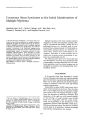
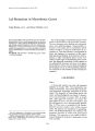
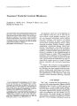
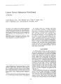
Show Journal of Clinical Neuro-ophthalmology 7(3):135--138, 1987. © 1987 Raven Press, Ltd., New York Cavernous Sinus Syndrome as the Initial Manifestation of Multiple Myeloma Sheridan Lam, M.D., Curtis E. Margo, M.D., Roy Beck, M.D., Thomas J. Pusateri, M.D., and Stephen Pascucci, M.D. A 76-year-old man developed a cavernous sinus syndrome as the initial manifestation of multiple myeloma. Although clinically the patient had stage IA disease, which is typically associated with a favorable response to therapy, his disease was rapidly fatal. This case emphasizes a weakness of the traditional staging system because it does not take into account certain clinical and histopathologic aspects of myeloma relevant to extramedullary plasmacytomas. Since disorders of ocular motility are more likely to be associated with extramedullary myeloma than myeloma confined to the marrow, clinicians need to be aware of the limitations in the clinical staging system and the potential problems associated with anaplastic plasmacytomas. Key Words: Cavernous Sinus-Multiple myelomaPlasmacytoma. From the Section of Ophthalmology, (S.L., C.E.M., R.B., T.J.P., S.P.), Veterans Administration Hospital, and the Departments of Ophthalmology (C.E.M., R.B.) and Pathology, (C.E.M.) University of South Florida, College of Medicine, Tampa, Florida. Address correspondence and reprint requests to Dr. S. Lam, Department of Ophthalmology, College of Medicine, Box 21, University of South Florida, 12901 North 30th Street, Tampa, FL 33612, U.s.A. 135 Multiple myeloma is the most common primary malignancy of bone and frequently presents clinically as a peripheral neuropathy, usually due to a pathologic fracture of a vertebral body or compression from a plasmacytoma (1). Intracranial extramedullary plasmacytomas are relatively unusual; thus, cranial nerve involvement is uncommon. Disorders of ocular motility due to orbital and cavernous sinus involvement in myeloma are exceptionally rare (2). The following case report of a 76-year-old man who developed a cavernous sinus syndrome from extramedullary myeloma illustrates how the traditional guidelines used for clinical staging may be unreliable for patients with predominately extramedullary disease. CASE REPORT A 76-year-old white man described a 3-week history of diplopia associated with right periorbital pain. He had been in good health previously, and there was no other pertinent past medical history except for bilateral visual loss from age-related macular degeneration. Examination revealed 20/400 visual acuity and a dense central scotoma in each eye. There was less than 1 mm of right ptosis and no proptosis. Pupils were equal in size and reacted normally to light. There was limitation of adduction, elevation, and depression of the right eye with normal motility on the left (Fig. 1). Large macular scars in both fundi were compatible with the visual acuity. There was hypesthesia in the distribution of the ophthalmic division of the right fifth cranial nerve. With forced duction testing, the right eye could be moved easily in all directions. General neurologic and systemic examinations were normal. 136 S. LAM ET AL. FIG. 1. The patient is exotropic and has a minimal amount of right ptosis in primary gaze. He is unable to adduct the right eye and has limitations of elevation and depression on the same side. Pertinent laboratory studies showed normal blood count and routine blood chemistries including serum protein concentration. Serologic tests for syphilis were negative, and a Westergren sedimentation rate was 90 mm/h. Urinalysis was 1+ positive for protein on dip stick. Analysis of cerebral spinal fluid (CSF) revealed a glucose concentration of 76 mg/dl and protein concentration of 25 mg/dl. Microscopic examination of the CSF showed three mature lymphocytes. Computed tomography (CT) and magnetic resonance scanning of the orbits and brain with special attention to the caverous sinus were normal. Despite treatment with oral prednisone, over a 2-week period the patient developed a complete ophthalmoplegia, complete ptosis, and a dilated, unreactive pupil of the right eye. Severe pain persisted. A 3-day course of 80 mg intravenous methylprednisolone per day had no salutary effects. The patient was discharged from the hospital with the diagnosis of cavernous sinus syndrome of unknown etiology. Three weeks later, the patient was readmitted because of right thigh pain. Radiologic studies showed a solitary osteolytic lesion in the shaft of the femur. Serum protein electrophoresis disclosed an abnormal monoclonal peak. Serum IgA -!, Mi,~r) was 776 mg/dl (normal, 50-250). "'re ~'resent in the urine. I Clill Nellrv-ophthalmol. Vol. 7. No.3. 1987 Open biopsy of the bone lesion showed an anaplastic plasmacytoma, which stained positive for IgA and K light chains using an immunofluorescent technique (Fig. 2). The tumor did not stain for IgG, IgM, or 'lI. light chains, indicating a monoclonal composition. CT of the extremities and trunk revealed no other bone lesions. A bone FIG. 2. Biopsy from the femur reveals a mass of anaplastic tumor cells that have destroyed bone trabeculae. The cells show little resemblance to normal plasma cells and could be mistaken for an undifferentiated lymphoma or a poorly differentiated carcinoma (hematoxylin and eosin, x 260). CAVERNOUS SINUS SYNDROME AND MYELOMA 137 marrow biopsy showed an increase in normal-appearing plasma cells (30%). The patient was staged clinically as IA (3). He received local radiation to the femur (2,250 rads total) and systemic chemotherapy (12 mg melphalan orally per day). During the next month, the patient's general condition deteriorated. He developed cardiac arrhythmias and progressive azotemia, and his mental function declined. Serum concentrations of IgA continued to rise. The patient died 3 months after the onset of his diplopia. AUTOPSY FINDINGS The patient had widespread myeloma involving both kidneys, thyroid gland, both adrenal glands, retroperitoneal soft tissue, heart, and the right cavernous sinus (Fig. 3). Visceral and soft-tissue tumors consisted of pleomorphic cells with irregularly shaped nuclei, coarse chromatin, and prominent nucleoli. Multiple bone marrow biopsies showed 30-40% plasmacytosis, but the marrow plasma cells appeared normal morphologically. The right cavernous sinus was totally filled with anaplastic plasma cells. Cranial nerves within the sinus were virtually replaced by tumor. DISCUSSION A clinical staging system for multiple myeloma was recommended in 1975 to help provide better initial assessment and follow-up for individual patients, and to lead to improved study design and analysis in therapeutic trials (3). Using multivariate regression analyses, Durie and Salmon (3) de-termined that myeloma cell mass could be accurately predicted based on four variables: (a) extent of bone lesions; (b) hemoglobin concentration; (c) serum calcium concentration; and (d) M-component concentration in serum and urine. Using these factors, patients with myeloma can be placed into three categories that correlated with low myeloma cell mass (Stage I), intermediate myeloma cell mass (Stage II), and high myeloma cell mass (Stage III). In addition, since prognosis worsens if renal dysfunction is present, each stage was subdivided into groups based on renal function tests (A, normal renal function; B, renal insufficiency). According to this staging system, our patient had Stage IA disease, which usually responds favorably to chemotherapy (78% of Stage I patients will have a complete response) (8). A case of anaplastic myeloma was reported by Okano and associates in 1966, under the rubric "plasmacytic reticulum cell sarcoma" (5). This atypical plasma cell tumor did not respond to therapy; at autopsy, it was shown to involve multiple visceral organs, soft tissue, and skin (5). Histologically, the cells were anaplastic. Electron microscopic findings were consistent with plasma cells, including elaborate rough-surfaced endoplasmic reticulum. There is reason to believe that anaplastic and poorly differentiated myelomas behave more aggressively than do well-differentiated tumors. A study of the extraskeletal spread of myeloma in 57 consecutively autopsied cases found two distinct patterns of dissemination: (a) tumors confined to adjoining paraskeletal tissue, and (b) tumors at distant sites (6). The most common sites of distant spread were spleen, liver, lymph nodes, and FIG. 3. The cavernous sinus is filled with tumor. Tumor has replaced the cranial nerves, but the carotid artery (C) is uninvolved. Only a minute portion of nerve located centrally in the sinus can still be identified (arrow) (hematoxylin and eosin, x 10). Inset: Tumor cells are undifferentiated and are similar to cells from the initial biopsy ( x 250). JClill Neuro-Ol'hthallllOI, Vol. 7, No.3, 1987 138 S. LAM ET AL. kidneys. Patients dying of distant spread had a higher proportion of poorly differentiated tumors than patients dying with skeletal and paraskeletal involvement. The large majority of extramedullary plasmacytomas are composed of cells that are moderately to well differentiated (4,7). Some can exist as solitary lesions for years without spreading. Helmus, in reviewing the world literature in 1964, found 147 cases of extramedullary plasmacytoma, 90% of which occurred in the upper respiratory tract and digestive tract; approximately 40% of patients died from their disease (8). Solitary plasmacytomas with the best prognosis are composed of relatively well-differentiated cells (9). The degree of cellular atypia in patients having so-called smoldering myeloma is relatively minimal compared with the anaplastic cells seen in our patient (9). Anaplastic myeloma can be confused with poorly differentiated carcinoma, immunoblastic lymphoma, and a variety of other undifferentiated tumors. Since the bone marrow may be minimally involved, anaplastic myeloma could be misdiagnosed unless appropriate immunologic studies are performed. Anaplastic myeloma tends to affect visceral organs and soft tissue to a greater extent than bone. Advanced extramedullary disease may not elevate serum calcium or suppress red blood cell production, because bone involvement can be minimal despite widespread visceral involvement (4). The criteria used for clinically staging skeletal myeloma, therefore, may not be applicable in some cases of extramedullary myeloma. Multiple myeloma accounts for less than 4% of all cases of cavernous sinus syndrome (10). While the orbit and cavernous sinus are uncommon sites of extramedullary plasmacytoma, the initial signs or symptoms of the disease may be related to infiltration of these areas (11). To our knowledge, only one other patient with myeloma presenting with an oculomotor palsy has been reported (12). This I Clill Nellro-ophthalmol. Vol. 7, No.3. 1987 case differed from ours in that the lesion represented localized spread from a skeletal lesion in the sphenoid sinus and was relatively well-differentiated histologically. Had the proteinuria in our patient been further investigated initially with urinary protein electrophoresis, the diagnosis of myeloma would have been established several weeks sooner. The clinical staging system for myeloma has several weaknesses, since its criteria were designed to assess bony involvement primarily and because it does not take into account the histologic grade of tumor. REFERENCES 1. Osserman EF. Plasma-cell myeloma. II. Clinical aspects. N Engl! Med 1959;261:1006-114. 2. Clark E. Cranial and intracranial myelomas. Brain 1977;77: 61-81. 3. Durie BGM, Salmon SE. A clinical staging system for multiple myeloma. Correlation of measured myeloma cell mass with presenting clinical features, response to treatment, and survival. Cancer 1975;36:842-54. 4. Wiltshaw E. The natural history of extramedullary plasmacytoma and its relation to solitary myeloma of bone and myelomatosis. Medicine 1976;55:217-38. 5. Okano H, Azar HA, Osserman EF. Plasma reticulum cell sarcoma. Case report with electron microscopic studies. Am! Clin Pathol 1966;46:546-55. 6. Pasmantier MW, Azar HA. Extraskeletal spread in multiple plasma cell myeloma. A review of 57 autopsied cases. Cancer 1969;23:167-74. 7. Azar HA. Pathology of multiple myeloma and related growths. In: Multiple myeloma and related disorders, vol. l. Azar HA, Potter M, eds. New York: Harper & Row, 1973: 1-85. 8. Helmus C. Extramedullary plasmacytoma of the head and neck. Laryngoscope 1964;74:553-9. 9. Kyle RA, Greipp PRo Smoldering multiple myeloma. N Engl! Med 1980;302:1347-9. 10. Thomas JE, Yass RE. The parasellar syndrome: problems in determining etiology. Mayo Clinic Proc 1970;45:617-23. 11. Rodman HI, Font RL. Orbital involvement in multiple myeloma. Review of the literature and report of three cases. Arch OphthalmoI1972;87:30-5. 12. Sundaresan N, Noronha A, Hirschauer J, Siqueira EB. Oculomotor palsy as initial manifestation of myeloma. !AMA 1977;238:2052-3. |