| OCR Text |
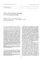
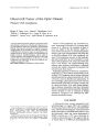
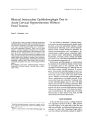
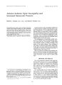
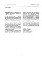
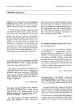
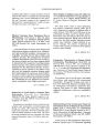
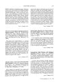
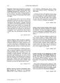
Show Joumal of Clmiml Nt'Jlro- 0I'" llw[ mo[ ogy 9(~): 1~ 6- 13U, 1989, Giant Cell Arteritis Associated with Progressive Systemic Sclerosis Saunders Lee Hupp, M. D. © 1989 Raven Press, Ltd" New York The clinical and autopsy findings in a patient with both systemic sclerosis ( scleroderma) and giant cell arteritis are presented. Giant cell arteritis and systemic sclerosis are autoimmune diseases affecting the elderly that may be associated with similar clinical symptoms of facial pain and arthralgias. Their coexistence is virtually unknown in the medical literature but is of great clinical importance. Systemic sclerosis is not treated with the steroid dosage necessary to prevent the morbid results of giant cell arteritis. An elevated sedimentation rate and/ or an abnormal temporal artery examination in an elderly patient with systemic sclerosis should alert the clinician to the possible coexistence of giant cell arteritis, Key Words: Giant cell arteritis- Systemic sclerosisTemporal arteritis- Scleroderma. From the Department of Ophthalmology, University of South Alabama College of Medicine, Mobile, Alabama. 1'., 1,1,, · '., t" rre" p', n, j,.' l1ce ,' nJ reprint "' quests to Dr. S, L. ; "" , c, ',', w" f: () phthalmology, Univl'rsity of South 126 Giant cell arteritis and systemic sclerosis are diseases of presumed autoimmune etiology that affect the elderly. Giant cell arteritis is associated with granulomatous vascular inflammation, often involving the vessels of the face ( temporal arteries) and orbit, that can result in ischemia, facial pain, and infarction. Systemic sclerosis is a multisystem disease characterized by inflammation, vasculitis, and deposition of fibrous tissue. The skin of the face and upper extremities can become atrophic and taut, resulting in " prune facies," facial pain, and limitation of movement of the temporomandibular joint. This report describes the clinical and pathologic findings of an elderly woman with long- standing systemic sclerosis who was found to have concomitant giant cell arteritis. CASE REPORT A 70- year- old white woman with known systemic sclerosis and open angle glaucoma complained of headaches, lethargy, and difficulty in chewing of increasing severity over a 2- month period. The glaucoma had been treated with Timoptic for several years with no progression of visual loss. She had no visual complaints. On examination her vision was 20/ 25 O. U. with normal ocular motility. Visual fields revealed bilateral nerve fiber bundle defects with more extensive loss in the left eye ( Fig. 1). The pupils were 3.0 mm, moderately reactive, with a slight afferent defect in the left eye. Intraocular tensions were 17 mm each eye. The optic discs were notable for early glaucomatous cupping ( Fig. 2). She had the typical " prune facies" associated with systemic sclerosis and rope- like pulseless temporal arteries ( Fig. 3). Hematocrit was 35% and a sedimentation rate ( Westergen) measured 53 mm/ h. Antinucleolar antibodies were present in a titer of 1: 40. The patient was hospitalized and started on oral prednisone 80 mg/ GIANT CELL ARTERITIS AND SYSTEMIC SCLEROSIS FIG. 1. Vision 20/ 25 O. U. Bilateral nerve fiber bundle defects are present with more extensive loss in left eye. 127 day. A temporal artery biopsy performed the next day showed extensive destruction of the artery wall, including the internal elastic lamina, by mononuclear and multinucleate giant cells consistent with giant cell arteritis ( Fig. 4). The patient had marked symptomatic improvement on steroids with the sedimentation rate decreased to 3 mm/ h. Five months later, while maintained on 30 mg/ day of prednisone, the patient experienced sudden abdominal pain and was found to have a ruptured sigmoid diverticulum. She developed peritonitis and suffered a pulmonary embolism. After a lengthy hospitalization she succumbed to generalized sepsis and respiratory failure. Pathological examination included a general autopsy as well as en bloc removal of the contents of both orbits. There was no vascular inflammation in either orbit. Serial sections of both intraocular and extraocular structures were normal for age. Only the temporal arteries showed extensive involvement with giant cell arteritis ( Fig. 4). Fibrosis typical of systemic sclerosis was identified in the lungs, kidneys, heart, and gastrointestinal tract ( Fig. 5). COMMENT Although systemic sclerosis and giant cell arteritis represent probable autoimmune- mediated dis- FIG. 2. Right and left optic nerves. Early glaucomatous changes are present with moderate media opacity from nuclear cataracts. 1Clill Nellro- ophthalmol. Vol. 9, No. 2. 1989 128 S. L. HUPP FIG. 3. Typical " prune facies" associated with systemic sclerosis. ease states, their coexistence is extremely rare and few documented cases appear in the literature ( 1,2). Both diseases affect the elderly and can be associated with head pain, arthritic complaints, and weight loss. Systemic sclerosis is a generalized disease of connective tissue involving the skin as well as extradermal organs, most often the gastrointestinal tract, lungs, heart, and kidneys ( 3). The disease affects females more often than males in a ratio of three to one, with an age at onset of ~ 40 years ( 4). Systemic sclerosis may involve only acral skin with limited extracutaneous involvement or may manifest as a rapidly progressive diffuse condition affecting trunk and extremities accompanied by marked inflammatory symptoms ( 5). The clinical manifestations of systemic sclerosis often begin in the fingers, hands, and face. Initially there is edema and swelling followed by hardening of the skin ( sclerosis), telangiectasias, and atrophy. The skin of the fingers may develop ulcers and calcific deposits. The taut facial skin results in a beaked nose, radial furrowing around the lips, and constricted opening of the mouth. Patients of- ". ,;, [ C() Jn both tl'mporoman- : !''' cd nerve com- J Clin Neuro- ophthalmol, Vol. 9, No. 2, 1989 - ~ .. ,.~ '~ .~.~ ........., FIG. 4. A: Left temporal artery biopsy. Note destruction of the muscular wall and internal elastic lamina by mononuclear and multinucleate giant cells. Extensive fibrinoid necrosis extends into the intima and the lumen is occluded by an organizing thrombus. x 1.2. B: Left temporal artery biopsy. Multinucleate foreign body giant cells in vessel wall. xa. pression from the surrounding sclerotic tissue ( 3). When extracutaneous involvement occurs, normal parenchymal tissue ( e. g., stomach wall, myocardium, lung) becomes replaced by sclerotic connective tissue. The final result in the pathogenesis of systemic sclerosis is fibrosis, but the initiating events remain obscure. Several initiating events have been proposed, including ( a) vascular endothelial damage leading to platelet aggregation and release of mediators of inflammation and fibroblast proliferation ( 6), ( b) disturbance in the regulation of connective tissue metabolism resulting in a subpopulation of " activated" fibroblasts ( 7), and ( c) altered immune response ( 8). There is considerable evidence that an altered immune state exists in systemic sclerosis ( 8) and giant cell arteritis ( 9- 11). The immune reaction in systemic sclerosis is typified by lymphohistiocytic infiltrations in the vicinity of small vessels early in the disease ( 3) and autoantibodies against nuclear and cellular antigens ( 12). Antinucleolar antibodies frequently present in patients with systemic sclero- GIANT CELL ARTERITIS AND SYSTEMIC SCLEROSIS 129 FIG. 5. Stomach wall. Note extensive fibrosis of the submucosa ( between arrows). Trichrome stain for collagen appears as black bands between arrows. Overlying mucosa contains postmortem autolytic changes. x 7. sis are rarely seen in other connective tissue diseases ( 13). The perivascular inflammatory reactions diminish or disappear later in the course of the disease ( 3). A perivascular infiltrate may also be seen in giant cell arteritis, but the main inflammatory reaction is directed against the internal elastic lamina ( 14). A reduction in OKT8 suppressor lymphocytes has been noted in both giant cell arteritis ( 9) and systemic sclerosis ( 8,15). These related immunologic changes present in both giant cell arteritis and systemic sclerosis suggest that their coexistence should not be unexpected. A specific treatment for systemic sclerosis is not known, but several drugs can reduce the clinical symptoms ( 3). Vasoactive agents such as plasma expanders, calcium channel blockers, and fibrinolytic agents are used in patients with acroscleroderma and Raynaud's disease ( 16,17). Antiinflammatory agents ( corticosteroids, cyclophosphamide, azathioprine) may improve the symptoms associated with myositis or interstitial lung involvement but do not seem to alter the progression of sclerosis ( 18). n- Penicillamine, an inhibitor of collagen biosynthesis, has been shown to reduce the cutaneous involvement of the disease ( 18). The detection of the presence of giant cell arteritis in a patient with systemic sclerosis is of major clinical importance. The treatment of systemic sclerosis is nonspecific and directed toward symptomatic improvement. It is likely not to include the high doses of steroids necessary to prevent the morbid complications, blindness and stroke, associated with giant cell arteritis. SUMMARY Giant cell arteritis and systemic sclerosis are the probable result of immunologic malfunction and it is reasonable that they may coexist in the same patient. This relationship is rarely noted possibly because the common clinical symptoms of giant cell arteritis, headache, proximal arthritis, and weight loss are often associated with systemic sclerosis. The diagnosis of systemic sclerosis is often evident from the clinical appearance, but the coexistence of giant cell arteritis may not be considered unless signs of ocular ischemia are present. Features strongly suggestive of giant cell arteritis include abnormal palpation ( enlargement, diminished pulses, or pain) of the temporal arteries and a markedly elevated sedimentation rate. If either of the above signs is present, steroid therapy should be initiated and a temporal artery biopsy performed. REFERENCES 1. Wyble M, Schimek RA. The simultaneous occurrence of two collagen diseases in the same patient. Trans Am Acad Ophthalmol Otolaryngol 1962; 66: 632- 41. 2. Perez- Jimenez F, Lopez- Rubio F, Canadillas F, JimenezAlonso). Jimenez- Pereperez J. Giant cell arteritis associated with progressive systemic sclerosis. Arthritis Rheum 1982; 25: 717- S. 3. Krieg T, Meurer M. Systemic scleroderma. JAm Acad Dermatol 1988; 18: 457- S1. 4. Medsger TA. Epidemiology of progressive systemic sclerosis. In: Black C, Meyers AR. eds. Systemic sclerosis ( scleroderma). New York: Gower, 1958: 53. 5. Rodnan GP, Jablonska S, Medsger TA. Classification and nomenclature of progressive systemic sclerosis ( scleroderma). Clin Rheum Dis 1976; 5: 5- 13. 6 Kahaleh MB, LeRoy EC. Endothelial injury in scleroderma- a protease mechanism. J Lilb Clin Med 1983; 101: 553-- 60. 7. Krieg T, Perlish IS, Mauch C, Fleischmajer R. Collagen synthesis by scleroderma fibroblasts. AmI NY Acad Sci 1986; 460: 375- S6. 8. Whiteside TL, Kumagai Y, Roumm AD, et al. Suppressor cell function and T lymphocyte subpopulations in periph- J Clil' Neuro- ophthalmol, Vol. 9, No. 2, 1989 130 S. L. HUPP eral blood of patients with progressive systemic sclerosis. Arthritis Rh~ lml 1983; 7: 841- 7. 9. Elling H, Elling P. Decreased level of suppressor/ cytotoxic T cells ( OKT8 + ) in polymyalgia rheumatica and temporal arteritis: relation to disease activity. I Rh~ lImatol 1985; 12: 306-- 9. to. Youinou PY, Pennec Y, Tande 0, Le Menn G. Immune complexes and autoantibodies in patients with giant cell arteritis and their relationship with autologous rosetteforming cells. Clill Exl' Rht'llmatol 1985; 3: 17- 21. 11. Ettlinger RE, Hunder GG, Ward LE. Polymyalgia rheumatica and giant cell arteritis. All/ llI R~ l' Med 1978; 29: 15- 22. 12. Tan EM, Rodnan GP, Gargia I. Moroi Y, Fritzler M]. Peebles C Diversity of antinuclear antibodies in progressive systemic sclerosis. Arthritis Rhellm 1980; 23: 617- 25. 13. Bernstein RM, Steigerwand Je. Tan EM. Association of an-tinuclear and antinucleolar antibodies in progressive systemic sclerosis. Clin Exp lmmunol 1982; 48: 43- 51. 14. Spencer WHo Ophthalmic pathology, an atlas and textbook, vol 3. Philadelphia: WB Saunders, 1986: 2377. 15. Keystone Ee. Lau C, Gladman DO, et al. Immunoregulatory T cell populations in patients with scleroderma using monoclonal antibodies. Clin Exp lmmunol 1982; 48: 443- 88. 16. Holti G. The effect of intermittent low molecular dextran infusion upon the digital circulation in systemic sclerosis. Br I Dermatol 1965; 77: 560- 8. 17. Kahan A, Weber S, Amar B, Saporta L, Hodara M. Nifedipine and Raynaud's phenomenon. Ann Intern Med 1981; 94: 546-- 52. 18. Steen VD, Medsger TA, Rodnan GP. o- Penicillamine therapy in progressive systemic sclerosis: a retrospective analysis. AIIII llltern Med 1982; 97: 652- 9. |