| OCR Text |
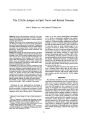
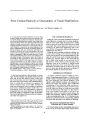
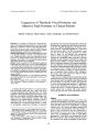
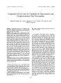
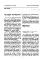
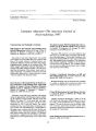
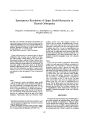
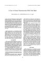
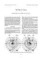
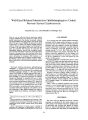
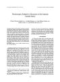
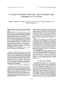
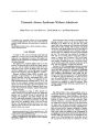
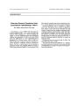
Show Journal of Neuro- Ophthalmology 19( 2): 148- 151, 1999. © 1999 Lippincott Williams & Wilkins, Inc., Philadelphia Traumatic Horner Syndrome Without Anhidrosis Shinji Oono, M. D., Isao Saito, M. D., Giichi Inukai, M. D., and Keizo Morisawa In a patient with a traumatic avulsion of the arm, magnetic resonance imaging showed the exact site of the lesion that produced Horner syndrome with preservation of sweating on the face. Key Words: Brachial avulsion- Horner syndrome- Miosis- Normal sweating- Ptosis. CASE REPORT On April 12, 1996, a 30- year- old man got his right arm caught in a conveyor belt, and the distal portion of the upper arm was amputated by traction. The patient was admitted to the Department of Orthopedics, Saga Medical School Hospital. The humerus was fractured slightly distal to its mid-portion. The latissimus dorsi and the teres major muscles were torn, and the middle and lower trunks of the brachial plexus were avulsed. In addition, his right upper eyelid was slightly ptotic, and the right pupil was smaller than the left, suggesting right- side Horner's syndrome. Magnetic resonance images ( MRIs) revealed cystic structures of approximately 1.0 cm in diameter, which showed isodensity with the cerebrospinal fluid at the spinal levels of C7 to Tl ( Fig. 1). A diagnosis of traumatic pseudomeningocele, reaching from C7 to Tl, was made. No other traumatic changes were detected in the spinal cord, and no neurologic signs and symptoms were observed except for the Horner syndrome already mentioned. The patient had emergency surgical stump plasty and free skin graft. Ocular examination on May 8, 1996, showed corrected visual acuity to be 20/ 20 in both eyes. Intraocular pressure was 15 mm Hg OD and 16 mm Hg OS. Eye position was orthophoric, and ocular motility was normal. The right pupil measured 4.0 mm and the left pupil 5.0 mm ( Fig. 2). Both pupils reacted briskly to light. The right eyelid was moderately ptotic, with good levator function. The palpebral fissures measured 7 mm on the right and 9 mm on the left. The ocular media were clear, and both fundi were normal. Manuscript received February 19, 1998; accepted February 17, 1999. From the Departments of Ophthalmology ( S. O., I. S., G. I.) and Orthopedics ( K. M.), Saga Medical School, Japan. Address correspondence and reprint requests to Shinji Oono, M. D., Department of Ophthalmology, Saga Medical School, Nabeshima, Saga 849- 8501, Japan. Sweat responses were recorded on bromphenol blue printing test paper which was devised by Sakurai and Montagna ( 1,2). Before the test, the skin was gently wiped with cotton moistened with absolute alcohol and then dried for a few minutes. The test paper was placed on the face, and sponge rubber was put on the paper. It was pressed gently but firmly by the examiner's hands so that the paper was diffusely and evenly pressed against the face. The sweat print was obtained on the paper as blue dots on an orange- yellow background approximately 2 minutes later. Sweating of the lower cheek, around the mouth and the jaw was examined with the same method. Sweating on the face was normal, including the medial aspect of the forehead and the side of the nose, and no asymmetric sweating was detected. On June 23, 1996, the pupillogram showed a normal constriction phase of the light reflex, but pupillary redi-lation after light exposure was incomplete, manifesting dilation lag ( 3). The diagnosis of Horner syndrome was thus confirmed. Pharmacologic pupil testing was performed. After bilateral instillation of 1.0% phenylephrine eye drops the right pupil dilated 2.0 mm ( from 4.0 to 6.0 mm), the normal left pupil only 0.5 mm ( from 5.0 to 5.5 mm), as shown in Figure 2. At the same time, the right-side ptosis was replaced by slight lid retraction. These findings suggested slight adrenergic supersensitivity on the injured side. DISCUSSION The efferent fibers of the oculosympathetic nerve innervate the superior cervical ganglion, which contains postganglionic neurons. These send fibers to the iris for pupillary dilation, to Muller's muscle for the lids, and ( in animals) to fibers for the nictitating membrane. Other neurons ( sudomotor, vasomotor, secretory, and pilomotor) supply their respective effectors. It has been known for many years that these functionally distinct neuron populations in the mammalian superior sympathetic ganglion are innervated by separate contingents of preganglionic fibers from the spinal cord. A degree of topical order exists within the ganglion in the arrangement of postganglionic neuron groups and between these and the location within the spinal cord of the preganglionic elements ( 4- 10). These findings have shown that most of the cord neurons for the pupil are contained in its first 148 HORNER SYNDROME WITHOUT ANHIDROSIS 149 V I ' • n ' i P l .* % ; ^ Off* if^ r i l- 1 _ | W^ TJ ' 1 vJ k 1 FIG. 1. Magnetic resonance images ( T1) show meningoceles at the spinal levels of C7 to T1, caused by avulsion of the ventral roots from the spinal cord. ( and second) thoracic segments, with their efferent fibers traveling to the sympathetic chain in the corresponding ventral roots. The upper and lower margins of the cord area vary to a degree among species and individual subjects. In man, also, the pupil fibers leave the spinal cord slightly rostral to the sudomotor and vasomotor fibers-that is, mostly by way of Tl, rather than T2 to T4, respectively ( 11- 14). Rare cases of pupil- sparing sympathetic defects have thus been described with all signs of Horner syndrome except for a perfectly normal pupil ( 10). The union of ventral spinal roots C5 to Tl forms the brachial plexus, which is located just distal to the scalene muscle. It consists of three trunks: upper ( C5, C6), middle ( C7), and lower ( C8, Tl). Lower plexus injury ( Klumpke type) causes segmental sensory and motor deficits including the regions innervated by the C8 and Tl spinal segments. It is well known that such injury is often accompanied by Horner syndrome, as was the case in our patient who also had avulsion of the seventh cervical ventral root. However, we are not aware of detailed descriptions of sudomotor function in the faces of such patients. Sudomotor loss appears to be the accepted consequence of such defects ( 12). It is commonly stated ( 15,16) that facial anhidrosis does not occur when Horner syndrome results from a lesion of the postganglionic sympathetic fibers. This is said to be because the sympathetic fibers to the eye travel with the internal carotid artery after leaving the superior cervical ganglion, whereas those destined for the sweat glands of the face accompany the external carotid artery ( 17). Even though our patient had a preganglionic lesion, he manifested the opposite of pupil- sparing Horner syndrome, in that he had deficits of pupillodilator and of Miiller's muscle innervation without anhidrosis in the face. Magnetic resonance images showed a pseudomen-ingocele from C7 to Tl, with complete nerve root avulsion from the spinal cord at these levels. The lesion undoubtedly caught most of the preganglionic pupillodilator and lid fibers to the superior sympathetic ganglion but left the more caudally located sudomotor and vasomotor FIG. 2. Top: Right- side Horner syndrome, with anisocoria and ptosis. Bottom: Anisocoria was reversed, and the ptosis disappeared after bilateral instillation of 1.0% phenylephrine, confirming the diagnosis of peripheral Horner syndrome. J Neuro- Ophlhalmol. Vol. 19, No. 2, 1999 150 S. OONO ETAL. Sudomotor fibers External carotid artery Superior cervical ganglion 1st Sudomotor fibers Oculosympathetic fibers Internal carotid artery Middle cervical ganglion Stellate ganglion - Inferior cervical ganglion - thoracic ganglion 2nd thoracic ganglion 3rd thoracic ganglion - 4th thoracic ganglion - White ramus PVR: Paravertebral route FIG. 3. Sympathetic pupillomotor and sudomotor fibers to the face ( modified after Morris et al. [ 13] and Palumbo [ 18]). Since the middle decades of this century, it has been generally accepted that most of the preganglionic sympathetic fibers arise from neurons in the lateral horns of the cervicothoracic spinal cord, with the pupillary neurons located rostrally, mostly in the Tl cord segment, and those for sudomotor function more caudally ( T2- T4). The sympathetic nerve fibers emerging from the cord in the ventral roots have always been assumed to travel for each segment with the rami communicantes to the respective paravertebral ganglia ( T1- T4), before turning cephalad to assemble in the cervical sympathetic chain. Palumbo ( 18) postulated that the pupillary fibers were not conducted by the rami communicantes but instead reached the stellate ganglion ( composed of the fused first thoracic and the inferior cervical ganglion) by a separated paravertebral route. The postganglionic oculosympathetic fibers for the pupil, the lids, and ( in animals) the nictitating membrane run from the superior cervical ganglion with the internal carotid plexus, but most of the facial sudomotor fibers travel by way of the external carotid plexus. In recent decades only a small area above the brow was said to have sudomotor deficiency in patients with internal carotid lesions. Recently, Morris et al. ( 13) stated that the sudomotor defect was more widespread, including the side of the nose and a larger portion of the head above the eye. elements intact. An explanation for this disparity has already been provided by the observations of Goetz ( 14). Using the techniques of stimulation and ablation of the exposed lower cervical and upper thoracic roots he was able to show that most of the pupillary fibers leave the spinal cord in the Tl ventral root. Fibers destined for the sweat glands of the face leave the spinal cord mainly in the T2 to T3 ventral roots. Hyperhidrosis of the face could be abolished in 90% of the patients without producing ptosis or miosis by resecting the T2 to T3 sympathetic ganglia. It is also noteworthy that Morris et al. ( 13) reported that none of their patients with Horner syndrome due to avulsion injuries of the roots of the branchial plexus ( C5 or C7- T1) had an abnormality of sweating. That sweating was spared in their patients as it was in ours was thus probably because the thoracic roots below Tl were not involved. These observations strongly suggest that the lesion between the Tl cord segment and its pathway to the sympathetic chain could cause Horner syndrome without anhidrosis, although it is substantially preganglionic. It is meaningful to reconsider the peripheral sympathetic pathways controlling the dilation of the pupil and sweating of the face in humans bearing in mind the works of Morris et al. ( 13) and of Palumbo ( 18). First, it is commonly accepted that the sudomotor fibers to the face ( except for a small area of the forehead) follow the external carotid artery after leaving the superior cervical ganglion. Morris et al. studied further details of this subject. They concluded that the sympathetic fibers for the sweat glands of the forehead and nose run with the internal carotid artery from which their blood supply also arises. Second, Palumbo postulated from his entire clinical experience that the preganglionic neurons controlling the pupil enter the upper portion of the stellate ganglion, the inferior cervical ganglion, by a separate paravertebral route after leaving the ventral roots of the eighth cervical and the first and/ or second thoracic nerves. Palumbo rejected the concept that these pathways pass through the first ramus communicans to the first thoracic ganglion. Taking these two statements in the literature into consideration, we have drawn a new diagram of the sympathetic pathway of the pupillomotor and the sudomotor fibers to the face ( Fig. 3). REFERENCES 1. Sakurai M, Montagna W. Experiments in the sweating on the palms of the green monkey. J Invest Dermatol 1965; 44: 87- 92. 2. Sakurai M. Use of bromphenol blue printing method for detecting sweat on the palm. J Hand Surg 1986; 11: 125- 30. 3. Pilley SFJ, Thompson HS. Pupillary " dilation lag" in Horner's syndrome. Br J Ophthalmol 1975; 59: 731- 5. 4. Langley JN. On the origin from the spinal cord of the cervical and upper thoracic sympathetic fibres, with some observations on the white and grey rami communicantes. Philos Trans R Soc Lond B BiolSci 1892; 183B: 85- 124. 5. Langley JN. On the regeneration of pre- ganglionic and of postganglionic visceral nerve fibres. J Physiol 1897; 22: 215- 31. 6. Murray JG, Thompson JW. The occurrence and function of collateral sprouting in the sympathetic nervous system of the cat. J Physiol 1957; 135: 133- 62. 7. Guth LG, Bernstein JJ. Selectivity in the re- establishment of synapses in the superior cervical sympathetic ganglion of the cat. Exp Neurol 1961; 4: 59- 69. 8. Naja A, Purves D. Specific innervation of guinea- pig superior cervical ganglion cells by preganglionic fibers arising from different levels of the spinal cord. J Physiol 1977; 264: 565- 83. 9. Ray BS, Hinsey JC, Hare K, Geohegan WA. Observations on the distribution of the sympathetic nerves to the pupil and upper extremity by stimulation of the anterior roots in man. Ann Surg 1943; 118: 647- 55. J Neuro- Ophthalmol, Vol. 19, No. 2, 1999 HORNER SYNDROME WITHOUT ANHIDROSIS 151 10. Loewenfeld IE. The pupil: Anatomy, physiology and clinical applications. Ames, LA: Iowa State University Press, 1993; Chap. 6,25: 327- 31,1147- 9; Figures 6- 7, 6- 8; Tables 6- 3, 6- 4, 6- 12. 11. Jaeger R, Whiteley WH. Avulsion of the brachial plexus. Report of six cases. JAMA 1953; 153: 633- 5. 12. Lister G. The hand: diagnosis and indications. 2nd ed. Edinburgh: Churchill Livingstone, 1984: 167- 8. 13. Morris JG, Lee J, Lim CL. Facial sweating in Horner's syndrome. Brain 1984; 107: 751- 8. 14. Goetz RH. The surgical physiology of the sympathetic nervous system with special reference to cardiovascular disorders, lnt Abstracts Surg 1948; 87: 417- 39. 15. Keane JR. Oculosympathetic paresis. Analysis of 100 hospitalized patients. Arch Neurol 1979; 36: 13- 15. 16. Jaffe NS. Location of lesions causing Horner's syndrome. Arch Ophthalmol 1950; 44: 710- 28. 17. Wilson WC. Observations relating to the innervation of the sweating glands of the face. Clin Sci 1936; 2: 273- 86. 18. Palumbo LT. A new concept of the sympathetic pathways to the eye. Ann Ophthalmol 1976; 8: 947- 54. J Neuro- Ophthalmol, Vol. 19, No. 2, 1999 |