| OCR Text |
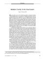
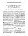
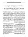
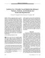
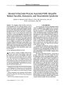
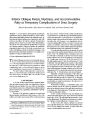
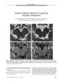
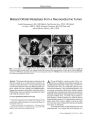
Show ORIGINAL CONTRIBUTION Bilateral Anterior Ischemic Optic Neuropathy After Liposuction Rod Foroozan, MD and Joseph Varon, MD, FACP, FCCP, FCCM Abstract: A 30- year- old woman had bilateral anterior ischemic optic neuropathy after undergoing large- volume liposuction. Visual function remained stable over a four-month follow- up, with decreased visual acuity and marked constriction of the visual fields. To our knowledge, this is the second reported case of ischemic optic neuropathy in this setting. ( JNeuro- Ophthalmol 2004; 24: 211- 213) Liposuction is one of the most commonly performed cosmetic procedures in the United States and is generally considered a safe procedure. However, serious complications, including fatalities, have been reported ( 1,2). We report a patient in whom visual loss from bilateral anterior ischemic optic neuropathy ( AION) developed after liposuction. This is the second reported case of AION after liposuction. CASE REPORT A 30- year- old woman with a history of obesity ( 218 lbs [ 99 Kg], body mass index > 35%) and hypothyroidism underwent high- volume tumescent liposuction and ab-dominoplasty. Preoperative blood pressure had been 121/ 77 mm Hg, pulse 79/ min, and hemoglobin 13.9 g/ dL. The 4.5- hour procedure was performed under general endotracheal anesthesia, during which 22,000 mL of fat was removed. Her postoperative weight loss was 65 pounds ( 29.5 kg). The intra- operative blood pressure was 120/ 50 mm Hg for nearly the entire procedure. She was given 12,100 mL of tumescent fluid, which included 3 mL of epinephrine 1: 1000 and 75 mL of 1% lidocaine, and 6700 mL of crystalloid fluid intravenously. In the recovery room, she reported blurred vision, attributed to the effects of general anesthesia. At the time of her visual symptom, blood pressure was 90/ 56 mm Hg, and Neuro- Ophthalmology Service ( RF), Cullen Eye Institute, Baylor College of Medicine, Houston, Texas, and The University of Texas Health Science Center ( JV), St. Luke's Episcopal Hospital, Houston, Texas. Address correspondence to Rod Foroozan, MD, Neuro- Ophthalmology Service, Cullen Eye Institute, Baylor College of Medicine, 6565 Fannin NC- 205, Houston, Texas, 77030; E- mail: foroozan@ bcm. tmc. edu pulse was 116/ min and regular. Vital signs remained essentially unchanged for the next 60 minutes. Systolic blood pressure remained between 100 and 110 mm Hg and diastolic blood pressure between 50 and 70 mm Hg for the remainder of the 24- hour hospital stay. Four days later, she developed headaches and dyspnea; six days after that, she came to the emergency department. Blood pressure was 110/ 60 mm Hg, pulse was 100/ min and regular, and respirations were 20/ min. Spiral computed tomography of the chest revealed multiple pulmonary emboli. Duplex ultrasonography of the lower extremities revealed no venous abnormalities. She had thrombocytopenia ( platelets 42,000/ mm3) and anemia ( hemoglobin 7.0 g/ dL, hematocrit 21.6%). She was transfused with 2 units of packed red blood cells and transferred to the critical care service, where anticoagulation with heparin was initiated for the pulmonary emboli. Ophthalmic examination revealed a near visual acuity of 20/ 70 OU. Pupils measured 8 mm in dim illumination and reacted sluggishly to light, with a left relative afferent pupillary defect. She was unable to identify any of the Ishi-hara pseudoisochromatic color plates with either eye and had inferior altitudinal visual field defects to confrontation. Bilateral pallid optic disc edema and hemorrhages were present within the retinal nerve fiber layer ( Fig. 1). The remainder of the fundus examination was normal. Magnetic resonance imaging and magnetic resonance venography of the brain revealed thrombosis of the right internal jugular vein, as well as the right transverse and sigmoid sinuses ( Fig. 2). Hemoglobin was 9.5 g/ dL and she received an additional 2 units of packed red blood cells. Lumbar puncture was not performed because of thrombocytopenia and the anticoagulation. Because of a concern that increased intracranial pressure might be present, she was started on acetazolamide, 500 mg twice daily. One week later, distance visual acuity was 20/ 60 OU and pupils still showed a left relative afferent pupillary defect. Automated perimetry revealed dense inferior altitudinal and superior arcuate visual field defects OU. Two weeks later, visual acuity was 20/ 40 OU with sectoral pallor of the inferior portions of the optic discs OU. Visual fields were unchanged. J Neuro- Ophthalmol, Vol. 24, No. 3, 2004 211 JNeuro- Ophthalmol, Vol. 24, No. 3, 2004 Foroozan and Varon FIG. 1. Bilateral pallid optic disc edema and nerve fiber layer hemorrhages associated with bilateral visual loss 11 days after high- volume liposuction. Four months after her initial examination, visual acuity was 20/ 50 OD and 20/ 60 OS with sluggishly reactive pupils and a left relative afferent pupillary defect. Automated perimetry revealed inferior altitudinal and superior arcuate visual field defects bilaterally ( Fig. 3). Both optic discs were pale. DISCUSSION High- volume liposuction (> 1500 mL of fat aspirated), as performed in this case, may be associated with a relatively high rate of morbidity and mortality ( 3). Pulmonary embolism, as occurred in our patient, is estimated to occur in 1: 2000 patients undergoing liposuction and has been implicated in 20% of fatalities ( 2). Other complications have included viscus perforation, fat embolism, cardiorespiratory failure, deep venous thrombosis, infection, and hemorrhage ( 2,3). Visual loss after liposuction is rare, with only one previous report ( 4). In that case, unilateral hypotensive anterior ischemic optic neuropathy occurred in a 47- year- old woman who underwent liposuction of the abdomen, thighs, and arms, and developed postoperative hypotension, tachycardia, and anemia ( 4). Visual loss developed OD on the second postoperative day and progressed to no light perception over the ensuing two days. Pallid optic disc edema was noted in the OD and the vision remained unchanged despite transfusion of 2 units of packed red blood cells. Magnetic resonance imaging and magnetic resonance angiography of the head were normal. Bilateral visual loss from AION after high- volume liposuction developed in our patient in association with pulmonary embolism and dural venous sinus thrombosis. The timing of her visual symptoms, decreased visual acuity, dyschromatopsia, visual field defects, and the appearance of the pallid swelling of the optic discs were characteristic of the perioperative optic nerve infarction that occurs in the setting of anemia and hypotension ( 5). Venous sinus thrombosis also developed, a complication not previously reported in this setting. The pulmonary embolism and dural venous sinus thrombosis may have resulted from rapid and severe dehydration. Although the earliest reports of liposuction recommended removal of no more than 1500 mL of fat, the introduction of epinephrine- containing wetting solutions has allowed larger volumes of fat to be aspirated. Removal of these larger volumes has generally been thought to be associated with an increased rate of complications, including fluid overload, pulmonary edema, lidocaine toxicity, deep venous thrombosis, and pulmonary embolism ( 6). Our patient underwent a large- volume removal of 22,000 mL of fat FIG. 2. Axial fluid attenuated inversion recovery ( FLAIR) magnetic resonance imaging of the brain ( A) reveals thrombosis within the right transverse sinus { arrow). Coronal contrast-enhanced T1- weighted magnetic resonance imaging ( B) shows clot within the right transverse sinus resulting in the " empty delta sign" { arrow). Magnetic resonance venography ( C) shows reduced or absent signal within the right internal jugular vein and the right transverse and sigmoid sinuses. 212 © 2004 Lippincott Williams & Wilkins Neuropathy After Liposuction JNeuro- Ophthalmol, Vol. 24, No. 3, 2004 FIG. 3. Automated perimetry ( Humphrey 24- 2) performed four months after liposuction shows inferior altitudinal and superior arcuate defects in both eyes. and was given 12,100 mL of tumescent fluid and 6700 mL of crystalloid fluid. These changes likely contributed to the ensuing hypotension and anemia that precipitated the bilateral AION. REFERENCES 1. Rao RB, Ely SF, Hoffman RS. Deaths related to liposuction. NEngl J Med 1999; 340: 1471- 5. 2. Grazer FM, de Jong RH. Fatal outcomes from liposuction: census survey of cosmetic surgeons. Plast Reconstr Surg 2000; 105: 436^ 18. 3. Albin R, de Campo T. Large- volume liposuction in 181 patients. Aesthetic Plast Surg 1999; 23: 5- 15. 4. Minagar A, Schatz NJ, Glaser JS. Liposuction and ischemic optic neuropathy. Case report and review of literature. J Neurol Sci 2000: 181: 132- 6. 5. Foroozan R, Buono LM, Savino PJ. Optic disc structure and shock-induced anterior ischemic optic neuropathy. Ophthalmology 2003: 110: 327- 31. 6. Commons GW, Halperin B, Chang CC. Large- volume liposuction: a review of 631 consecutive cases over 12 years. Plast Reconstr Surg 2001; 108: 1753- 63. 213 |