| OCR Text |
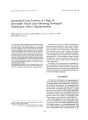
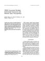
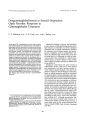
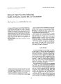
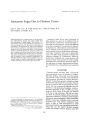
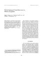
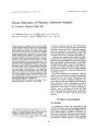
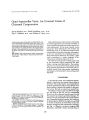
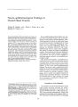
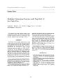
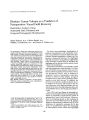
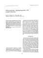
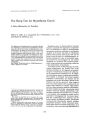
Show ! ourtlal of ClitJical Ne, mrophthaimoiosy 11( 4): 284- 287, 1991. Orbital Ischemia, Ophthalmoparesis, and Carotid Dissection Steven L. Galetta, M. D., Alan Leahey, M. D., Charles W. Nichols, M. D., and Eric C. Raps, M. D. © 1991 Raven Press, Ltd., New York We report a patient who developed an acute loss of vision in the left eye associated with proptosis, ophthalmoparesis, conjunctival injection, and chemosis. Funduscopy revealed optic disc swelling, and retinal whitening consistent with an ophthalmic artery occlusion. Angiography disclosed bilateral carotid dissections presumably resulting from head trauma 11 days earlier. An orbital ischemic syndrome may be a delayed manifestation of traumatic carotid dissection and precede cerebral hypoperfusion. Key Words: Orbital ischemia- Carotid dissectionOphthalmoparesis. From the Departments of Neurology ( S. L. G., E. eR.) and Ophthalmology ( S. L. G., A. L., eW. N.), Hospital of the University of Pennsylvania, Philadelphia, Pennsylvania, U. S. A. Address correspondence and reprint requests to Dr. Steven L. Galetta at Department of Neurology, Hospital of the Univer< ity of Pl'nnwlv, mia. 14110 Spruce Street, Philadelphia, fl ~ ~ ~ 1 {\ ~ I I S A 284 Dissection of the carotid artery results when intraluminal blood enters the arterial wall and separates its component layers. Arterial dissections may be spontaneous or secondary to minor trauma; others result from obvious blunt and penetrating trauma to the head and neck ( 1). Neuroophthalmic complications of carotid dissection include amaurosis fugax ( 1), ischemic optic neuropathy ( 2,3), central retinal artery occlusion ( 3,4), Horner's syndrome ( 5), homonymous hemianopsia ( 6), abducens nerve palsy ( 7), and an ocular ischemic syndrome ( 8). Oculosympathetic paralysis is the most common neuro- ophthalmologic finding in carotid dissection; it occurs in approximately 50% of patients ( 1,9). We report a patient who developed orbital ischemia and ophthalmoparesis following bilateral traumatic carotid dissection. CASE HISTORY Eleven days after an automobile accident, a 48year- old man presented to the Scheie Eye Institute with a 4- hour history of painless visual loss in the left eye. Examination disclosed 20/ 60 acuity in the right eye and no light perception in the left eye. The pupils were equal in size, with a large left afferent pupillary defect. Ocular motility was full and slit examination showed mild injection of the left conjunctiva. Ophthalmoscopy disclosed diffuse retinal whitening, with a cherry red spot in the left fovea. The right fundus appeared normal. He was treated with Diamox, ocular massage, Timoptic, and anterior chamber paracentesis without visual improvement. Three hours later, left eye abduction and adduction were limited to 90% of normal. Supraduction and infraduction were 70% of normal. Forced duction testing showed no restriction and facial sen- ORBITAL ISCHEMIA 285 sation was intact. He had 3 mm of left proptosis and mild chemosis. Funduscopy now disclosed a diffusely ischemic retina with an elevated band of retinal whitening extending from the edge of the swollen optic nerve head to the temporal border of the fovea ( Fig. 1). The patient was transferred to the University of Pennsylvania Medical Center for a cerebral arteriogram that revealed bilateral carotid dissections at the bifurcation of the common carotid artery. The left internal carotid was totally occluded and the left ophthalmic artery filled via the external carotid artery and meningeal branches ( Fig. 2). Several hours later, the patient developed intermittent left lower extremity weakness, and a left neglect. Intravenous Heparin was started and a routine MRI with gadolinium contrast showed an occluded left internal carotid with a patent cavernous sinus and normal ophthalmic veins ( Fig. 3). On T2- weighted images, increased signal intensity was found in the left and right parietal watershed regions. One week into the patient's hospitalization, new choroidal infarcts were detected in the right macula region. Three days later, his visual acuity was 201 40 in the right eye and NLP in the left eye. The left eye proptosis had resolved and ocular motility was normal. Two months after discharge and while on warfarin therapy, his visual acuity in the right eye was 20/ 20 with a normal fundus. There was no light perception vision in the left eye, and ophthalmoscopy showed a pale gliotic optic nerve with sheathing and narrowing of the retinal vessels. He had a mild left facial droop and ipsilateral hyperreflexia. FIG. 1. Fundus photograph of left eye demonstrating elevated band of retinal whitening in macula region ( black arrow) and optic nerve head swelling. There is embolic material in the superior temporal arcade ( white arrow). FIG. 2. Left carotid angiogram shows a tapering occlusion consistent with an arterial dissection. DISCUSSION Carotid dissection accounts for at least 5% of cerebral ischemic episodes in young adults ( 10). Abrupt facial, ear, or neck pain usually signifies the onset of the dissection and may precede symptomatic ocular or cerebral ischemia by hours to days ( 1). About 5- 10% of carotid dissections are bilateral ( 1). A variety of factors have been implicated in the pathogenesis of carotid dissection, including fibromuscular dysplasia, cystic medial necrosis, syphilis, pharyngeal infection, extension of aortic dissection, artheromatous disease, cerebral aneurysm, and trauma ( 1,9). The natural history of carotid dissection remains unclear, but cerebral and ocular ischemic symptoms are usually delayed for several hours to days following the initial trauma ( 1). Recently, attention has been drawn to the ophthalmic vaso- occlusive complications of carotid dissection including anterior and posterior ischemic optic neuropathy ( 2,3), an ocular ischemic syndrome ( 8), and central retinal artery occlusion ( 3,4) ( Table 1). The mechanism of ocular ischemia in these patients may be the result of either hypoperfusion or embolization ( 3). Our patient had embolic material in the superior retinal vessels. Orbital ischemia was manifested in our patient by proptosis, motility restriction, conjunctival injection, chemosis, optic disc swelling, and retinal whitening. This orbital ischemic syndrome preceded the onset of symptomatic cerebral ischemia by several hours. A traumatic carotid cavernous fistula was also considered, but the absence of a bruit and arterialization of conjunctival vessels made such a process unlikely. Angiography dem- JClin Neuro- ophthalmol. Vol. 11, No. 4, 1991 286 5. L. GALETTA ET AL. REFERENCES TABLE 1. Neuro- ophthalmic complications of carotid dissection patient with an isolated sixth nerve palsy secondary to a carotid artery dissection ( 7). The authors postulated that the abducens nerve palsy was either caused by an expanded intracavernous carotid wall or from disruption of the nerves blood supply. The clinical findings in our patient suggest extraocular muscle ischemia as another mechanism for ophthalmoparesis in carotid dissection. Recently, Wilson has documented three patients with transient ocular motor paresis associated with carotid artery occlusion ( 11). As suggested by Newman et al. ( 3), these three patients had no vascular risk factors and the primary etiology of the carotid occlusion may have been a dissection. Regardless of the underlying mechanism of a carotid occlusion, a transient unilateral ophthalmoparesis may herald its onset. Management options for carotid dissection include anticoagulation, antiplatelet agents, superficial temporal artery to middle cerebral artery anastomosis, direct exploration of the dissected internal carotid artery, and observation ( 9). Mokri et al. found complete recovery in 85% of their patients, regardless of the treatment modality chosen ( 9). Our patient improved with anticoagulation, volume expansion and hemodilution. Decisions regarding therapy should be individualized, particularly when a patient develops progressive neurologic signs despite expectant management. Reference ( 1,4,6,11 ) ( 3,4) ( 2) ( 4,6) ( 3) ( present case) ( 7) ( 5) ( 8) ( present case) ( 6) ( 6,12) Complication Amaurosis fugax Anterior ischemic optic neuropathy Posterior ischemic optic neuropathy Central retinal artery occlusion Ophthalmic artery occlusion Transient ophthalmoparesis Sixth nerve palsy Horner's syndrome ( postganglionic) Ocular ischemic syndrome Orbital ischemic syndrome Homonymous field defects Monocular or binocular scintillations A B FIG. 3. A: T2- weighted axial MR image showing high signal abnormality in lumen of left cavernous carotid consistent with proximal carotid occlusion ( black arrow). B: T2- weighted axial image of parietal region demonstrating high signal abnormality of watershed ischemia. onstrated bilateral carotid dissections without evidence of fistula. Our patient also had transient ophthalmoparesis as a manifestation of his carotid occlusion and arterial dissection. Maitland et aI. have desaibed a 1. Hart RG, Easton JD. Dissections of cervical and cerebral arteries. Neurol Clin 1983; 1: 155- 82. 2, Rivkin MJ. Hedges TR III, Logigian EL. Carotid dissection presenting as posterior ischemic optic neuropathy. Neurology 1990; 40: 1469. 3. Newman NJ. Kline LB, Leifer D, Lessell S. Ocular stroke and carotid artery dissection. Neurology 1989; 39: 1462- 4. 4. Bogousslavsky J. Despland PA. Regli F. Spontaneous ca- ORBITAL ISCHEMIA 287 rotid dissection with acute stroke. Arch Neural 1987; 44: 13740. 5. Kline LB, Vitek JJ, Raymon BC Painful Horner's syndrome due to spontaneous carotid artery dissection. Ophthalmology 1987; 94: 22&- 30. 6. Fisher CM, Ojemann RG, Roberson GH. Spontaneous dissection of cervico- cerebral arteries. Call JNeural Sci 1978; 5: 9- 19. 7. Maitland CG, Black JL, Smith WA, Abducens nerve palsy due to spontaneous dissection of the internal carotid artery. Arch Neural 1983; 40: 44S- 9. 8. Duker JS, Belmont JB. Ocular ischemic syndrome second-ary to carotid artery dissection. Am J Ophthalmol 1988; 106: 750- 2. 9. Mokri B, Sundt TM, Houser OW, Piepgras DG. Spontaneous dissection of the cervical internal carotid artery. Ann Neural 1986; 19: 12&- 38. 10. Hart RG, Easton JD: Dissections. Stroke 1985; 16: 925. 11. Wilson WB, Leavengood JM, Ringel SP, Bott AD. Transient ocular motor paresis associated with acute internal carotid artery occlusion. Ann Neural 1989; 25: 28&- 90. 12. Ramadan NM, Tietjen GE, Levine SR, Welch KMA. Scintillating scotomata associated with internal carotid artery dissection: report of three cases. Neurology 1991; 41: 1084- 7. I Clin NeuTo- ophthalmol, Vol. 11. No. 4, 1991 [MKorbitalischemicsyndrome] |