John A. Moran Eye Center Neuro-Ophthalmology Collection: A variety of lectures, videos and images relating to topics in Neuro-Ophthalmology created by faculty at the Moran Eye Center, University of Utah, in Salt Lake City.
NOVEL: https://novel.utah.edu/
TO
Filters: Collection: ehsl_novel_jmec
1 - 25 of 4
| Identifier | Title | Description | Subject | ||
|---|---|---|---|---|---|
| 1 |
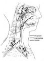 |
Figure-04 | Anatomy of the Oculosympathetic Pathway | Anatomy of the oculosympathetic pathway. (Maloney WF, Younge BR, Moyer NJ: Evaluation of the causes and accuracy of pharmacologic localization in Horner's syndrome. Am J Ophthalmol 1980;90:394-402, Ophthalmic Publishing Company with permission.) | Anatomy of the Oculosympathetic Pathway; Horner's Syndrome |
| 2 |
 |
1-2 | Dilation Lag | Two examples of dilation lag (Horner's syndrome). In the first example, the right pupil dilates much faster than the left pupil when the light is turned out. In the second example, the left pupil dilates much faster than the right pupil when the light is turned out. Discussion of methods of document... | Dilation Lag; Horner Syndrome; Horner's Syndrome |
| 3 |
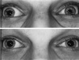 |
Figure-21 | Left-sided Dilation Lag in a Man with Horner's Syndrome | Left-sided dilation lag in a 29-year-old man with Horner's syndrome caused by a posterior mediastinal ganglioneuroma. Note that the degree of anisocoria is greater after 5 seconds in darkness (top) compared with findings after 15 seconds in darkness (bottom). | Diagnosis, Horner Syndrome; Physiopathology, Horner Syndrome; Reflex, Pupillary; Dilation Lag; Horner's Syndrome |
| 4 |
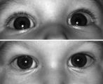 |
Figure-22 | Right-sided Pseudo-Horner's Syndrome | Right-sided pseudo-Horner's syndrome in an 8-month-old infant referred because her mother had noted a larger pupil on the left for a few months and her pediatrician thought the right upper lid was droopy. Both pupils reacted normally to light and darkness, the degree of anisocoria was similar in bot... | Infant; Diagnosis, Horner Syndrome; Etiology, Horner Syndrome; Horner's Syndrome |
1 - 25 of 4