AAO-NANOS Neuro-Ophthalmology Clinical Collection: Derived from the AAO-NANOS Clinical Neuro-Ophthalmology collection produced on CD. The images are of selected cases from the NANOS teaching slide exchange, and the CD was produced under the direction of Larry Frohman, MD and Andrew Lee, MD.
The American Academy of Ophthalmology (AAO); The North American Neuro-Ophthalmology Association (NANOS).
NOVEL: https://novel.utah.edu/
TO
Filters: Collection: ehsl_novel_aao_nanos
1 - 25 of 6
| Title | Description | Subject | ||
|---|---|---|---|---|
| 1 |
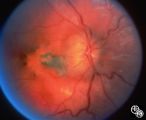 |
Isolated Congenital Optic Disc Anomalies | This 8-year-old boy presented with a 2-week history of decreased vision in the right eye. He had undergone a normal MRI and CSF examination, including intracranial pressure, before neuro-ophthalmologic assessment. The fundus photographs and fluorescein angiograms show subretinal neovascularization a... | Pseudopapilledema; Edema; Papilledema; Retinal Neurovascularization |
| 2 |
 |
Isolated Congenital Optic Disc Anomalies | This 8-year-old boy presented with a 2-week history of decreased vision in the right eye. He had undergone a normal MRI and CSF examination, including intracranial pressure, before neuro-ophthalmologic assessment. The fundus photographs and fluorescein angiograms show subretinal neovascularization a... | Pseudopapilledema; Edema; Papilledema; Retinal Neurovascularization |
| 3 |
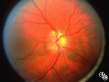 |
Isolated Congenital Optic Disc Anomalies | This image shows drusen that are especially prominent superotemporally. Pair with 92_64 and 92_67. | Optic Disc Drusen; Optic Nerve Drusen; Pseudopapilledema |
| 4 |
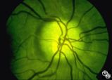 |
Isolated Congenital Optic Disc Anomalies | This optic disc displays multiple drusen. Note the pseudopapilledema here. One can differentiate this from true papilledema in that there is no obscuration of the vessel by the peripapillary nerve fiber layer as they cross the disc margin. This photograph was taken with barrier filters in place, but... | Optic Disc Drusen; Optic Nerve Drusen; Pseudopapilledema |
| 5 |
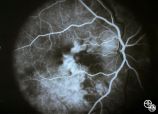 |
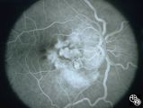
Pseudopapilledema With Retinal Neovascularization, Fluorescein Angiogram | This 8-year-old boy presented with a 2-week history of decreased vision in the right eye. He had undergone a normal MRI and CSF examination, including intracranial pressure, before neuro-ophthalmologic assessment. The fundus photographs and fluorescein angiograms show subretinal neovascularization a... | Pseudopapilledema; Edema; Papilledema; Retinal Neurovascularization; Fluorescein Angiogram |
| 6 |
 |
Pseudopapilledema With Subretinal Neovascularization on Fluorescein Angiogram | This 8-year-old boy presented with a 2-week history of decreased vision in the right eye. He had undergone a normal MRI and CSF examination, including intracranial pressure, before neuro-ophthalmologic assessment. The fundus photographs and fluorescein angiograms show subretinal neovascularization a... | Pseudopapilledema; Edema; Papilledema; Retinal Neurovascularization; Fluorescein Angiogram |
1 - 25 of 6