AAO-NANOS Neuro-Ophthalmology Clinical Collection: Derived from the AAO-NANOS Clinical Neuro-Ophthalmology collection produced on CD. The images are of selected cases from the NANOS teaching slide exchange, and the CD was produced under the direction of Larry Frohman, MD and Andrew Lee, MD.
The American Academy of Ophthalmology (AAO); The North American Neuro-Ophthalmology Association (NANOS).
NOVEL: https://novel.utah.edu/
TO
Filters: Collection: ehsl_novel_aao_nanos
1 - 25 of 21
| Title | Description | Subject | ||
|---|---|---|---|---|
| 1 |
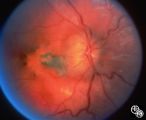 |
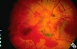
Isolated Congenital Optic Disc Anomalies | This 8-year-old boy presented with a 2-week history of decreased vision in the right eye. He had undergone a normal MRI and CSF examination, including intracranial pressure, before neuro-ophthalmologic assessment. The fundus photographs and fluorescein angiograms show subretinal neovascularization a... | Pseudopapilledema; Edema; Papilledema; Retinal Neurovascularization |
| 2 |
 |
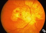
Isolated Congenital Optic Disc Anomalies | This 8-year-old boy presented with a 2-week history of decreased vision in the right eye. He had undergone a normal MRI and CSF examination, including intracranial pressure, before neuro-ophthalmologic assessment. The fundus photographs and fluorescein angiograms show subretinal neovascularization a... | Pseudopapilledema; Edema; Papilledema; Retinal Neurovascularization |
| 3 |
 |
Isolated Optic Neuritis/Neuropathy | Papilledema may produce visual loss due to chronic atrophic papilledema, secondary macular hemorrhage, exudate or edema, secondary ischemic optic neuropathy, or secondary subretinal neovascular membrane formation. Patients with papilledema and visual loss should be suspected of harboring one of thes... | Pseudotumor Cerebri/Papilledema; Edema |
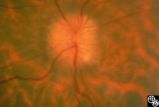
| 4 |
 |
Isolated Optic Neuritis/Neuropathy | Papilledema may produce visual loss due to chronic atrophic papilledema, secondary macular hemorrhage, exudate or edema, secondary ischemic optic neuropathy, or secondary subretinal neovascular membrane formation. Patients with papilledema and visual loss should be suspected of harboring one of thes... | Pseudotumor Cerebri/Papilledema; Edema |
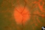
| 5 |
 |
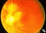
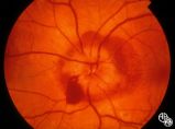
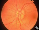
Isolated Optic Neuritis/Neuropathy | This 48-year-old man presented with a 1-month history of headache. Both discs had the appearance seen in this image, with prominent peripapillary nerve fiber layer myelination; the disc itself is hyperemic, with dilated, telangiectatic surface vasculature, suggesting true disc edema as well. | Pseudotumor Cerebri/Papilledema; Edema |
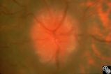
| 6 |
 |
Isolated Optic Neuritis/Neuropathy | Papilledema is a term reserved for optic disc edema related to increased intracranial pressure (eg. Papilledema, sixth nerve palsy, headache), a normal neuroimaging study, and an elevated opening pressure with normal cerebrospinal fluid contents. | Pseudotumor Cerebri/Papilledema; Edema |
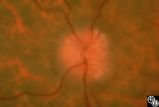
| 7 |
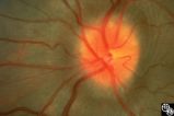 |
Isolated Optic Neuritis/Neuropathy | This image demonstrates Paton's lines in a 34-year-old patient with pseudotumor cerebri and chronic papilledema. | Pseudotumor Cerebri/Papilledema; Edema |
| 8 |
 |
Isolated Optic Neuritis/Neuropathy | This 42-year-old male with pseudotumor cerebri and chronic papilledema demonstrated refractile bodies, which can be seen with chronic optic disc edema. This image shows the chronic papilledema at presentation, with associated refractile hyaline bodies at the disc periphery in both eyes. Pair with 96... | Pseudotumor Cerebri/Papilledema; Edema |
| 9 |
 |
Isolated Optic Neuritis/Neuropathy | This 42-year-old male with pseudotumor cerebri and chronic papilledema demonstrated refractile bodies, which can be seen with chronic optic disc edema. This image shows the chronic papilledema at presentation, with associated refractile hyaline bodies at the disc periphery in both eyes. Pair with 96... | Pseudotumor Cerebri/Papilledema; Edema |
| 10 |
 |
Isolated Optic Neuritis/Neuropathy | This 42-year-old male with pseudotumor cerebri and chronic papilledema demonstrated refractile bodies, which can been seen with chronic optic disc edema. This image exhibits decreased disc edema and resolution of the refractile bodies OD after therapy. Pair with 96_01, 96_02, 96_04, 96_05, and 96_06... | Pseudotumor Cerebri/Papilledema; Edema |
| 11 |
 |
Isolated Optic Neuritis/Neuropathy | This 42-year-old male with pseudotumor cerebri and chronic papilledema demonstrated refractile bodies, which can be seen with chronic optic disc edema. This image exhibits decreased disc edema and resolution of the refractile bodies OD after therapy. Pair with 96_01, 96_02, 96_03, 96_05, and 96_06. | Pseudotumor Cerebri/Papilledema; Edema |
| 12 |
 |
Isolated Optic Neuritis/Neuropathy | This 42-year-old male with pseudotumor cerebri and chronic papilledema demonstrated refractile bodies, which can been seen with chronic optic disc edema. This image demonstrates later recurrence of the refractile bodies with worsening papilledema OD. Pair with 96_01, 96_02, 96_03, 96_04, and 96_06. | Pseudotumor Cerebri/Papilledema; Edema |
| 13 |
 |
Isolated Optic Neuritis/Neuropathy | This 42-year-old male with pseudotumor cerebri and chronic papilledema demonstrated refractile bodies, which can be seen with chronic optic disc edema. This image demonstrates later recurrence of the refractile bodies with worsening papilledema OD. Pair with 96_01, 96_02, 96_03, 96_04, and 96_05. | Pseudotumor Cerebri/Papilledema; Edema |
| 14 |
 |
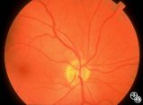
Isolated Optic Neuritis/Neuropathy | Papilledema in pseudotumor cerebri may result in adjacent choroidal or retinal folds. | Pseudotumor Cerebri/Papilledema; Edema |
| 15 |
 |
Isolated Optic Neuritis/Neuropathy | Papilledema may produce visual loss due to chronic atrophic papilledema, secondary macular hemorrhage, exudate or edema, secondary ischemic optic neuropathy, or secondary subretinal neovascular membrane formation. | Pseudotumor Cerebri/Papilledema; Edema |
| 16 |
 |
Isolated Optic Neuritis/Neuropathy | Papilledema is a term reserved for optic disc edema related to increased intracranial pressure. Fluid within the optic nerve sheath or elevation of the intraocular optic nerve head may be visible on magnetic resonance imaging studies of the head and orbit. | Pseudotumor Cerebri/Papilledema; Edema |
| 17 |
 |
Isolated Optic Neuritis/Neuropathy | Papilledema usually results in bilateral optic disc edema without visual loss. The blind spot may enlarge initially, but progressive visual field loss may occur with chronic optic disc edema. Asymmetric or frankly unilateral optic disc edema may occur due to structural disc fractures that prevent th... | Pseudotumor Cerebri/Papilledema; Edema |
| 18 |
 |
Isolated Optic Neuritis/Neuropathy | Papilledema usually results in bilateral optic disc edema without visual loss. The blind spot may enlarge initially, but progressive visual field loss may occur with chronic optic disc edema. Asymmetric or frankly unilateral optic disc edema may occur due to structural disc fractures that prevent th... | Pseudotumor Cerebri/Papilledema; Edema |
| 19 |
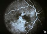 |
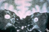
Pseudopapilledema With Retinal Neovascularization, Fluorescein Angiogram | This 8-year-old boy presented with a 2-week history of decreased vision in the right eye. He had undergone a normal MRI and CSF examination, including intracranial pressure, before neuro-ophthalmologic assessment. The fundus photographs and fluorescein angiograms show subretinal neovascularization a... | Pseudopapilledema; Edema; Papilledema; Retinal Neurovascularization; Fluorescein Angiogram |
| 20 |
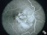 |
Pseudopapilledema With Subretinal Neovascularization on Fluorescein Angiogram | This 8-year-old boy presented with a 2-week history of decreased vision in the right eye. He had undergone a normal MRI and CSF examination, including intracranial pressure, before neuro-ophthalmologic assessment. The fundus photographs and fluorescein angiograms show subretinal neovascularization a... | Pseudopapilledema; Edema; Papilledema; Retinal Neurovascularization; Fluorescein Angiogram |
| 21 |
 |
Systemic Disorders With Optic Nerve and Retinal Findings | A 29-year-old African American woman presented with headaches, bilateral transient visual obscurations, blurred vision, numbness, and weakness of the lower extremities with myalgia and joint pains. She had an unplanned 12-pound weight loss over 2 months. A neurologist and internist diagnosed her wit... | Sarcoidosis; Papilledema |
1 - 25 of 21