John A. Moran Eye Center Neuro-Ophthalmology Collection: A variety of lectures, videos and images relating to topics in Neuro-Ophthalmology created by faculty at the Moran Eye Center, University of Utah, in Salt Lake City.
NOVEL: https://novel.utah.edu/
TO
Filters: Collection: ehsl_novel_jmec
| Identifier | Title | Description | Subject | ||
|---|---|---|---|---|---|
| 201 |
 |
2-19 | Superior Oblique Myokymia | Example of patients with superior oblique myokymia, a saccadic intrusion. First patient is seen to have intermittent, intorting movements with superimposed slight vertical deviations in right eye. Discussion of disorder as benign, but frequently disabling, as patients experience episodes of diplopia... | Superior Oblique Myokymia; Third Nerve Palsy |
| 202 |
 |
NOVEL_Moran_2-25 | Superior Oblique Myokymia | Close-up video of a patient with superior oblique myokymia (no audio.) | Superior Oblique Myokymia; Myokymia |
| 203 |
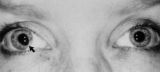 |
Figure-28 | Tadpole-shaped Pupil | Tadpole-shaped pupil in a 20-year-old women with frequent episodes of blurred vision and achiness of the right eye lasting several minutes. The patient took a photograph of her eyes during an attack to document the peaked, segmental dilation of her right pupil (black arow). (Thompson HS, Zackon DH, ... | Adult; Female; Humans; Complications, Iris Diseases; Diagnosis, Iris Diseases; Physiopathology, Iris Diseases; Pupil; Complications, Spasm; Diagnosis, Spasm; Physiopathology, Spasm; Tadpole Pupil |
| 204 |
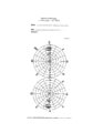 |
Tangent_screen_recording_chart | Tangent Screen Recording Chart | The tangent screen recording chart. | Tangent Screen |
| 205 |
 |
Tangent_Screen_Testing_Visual_Field | Tangent Screen Testing Visual Field | Description of tangent screen testing. | Tangent Screen; Visual Fields |
| 206 |
 |
Test Duane | Test Duane | ||
| 207 |
 |
Visual_Fields | Testing the Visual Fields | Demonstration of various methods of testing visual fields, including counting fingers, motion, and color of several objects. | Visual Fields; Examination, Ocular; Visual Field Loss |
| 208 |
 |
The_3_Step_Test_Digre.pdf | The 3 Step Test: Looking for a 4th Nerve Palsy | Description of the three step test (3 step test) used when looking for a 4th nerve palsy. | 3 Step Test |
| 209 |
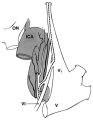 |
Figure-05 | The Course of the Postganglionic Segment of the Oculosympathetic Fibers from the Internal Carotid Artery | The course of the postganglionic segment of the oculosympathetic fibers from the internal carotid artery (ICA) to the orbit is depicted as a dotted line. Note that they briefly join the abducens nerve (cranial nerve VI) before joining the nasociliary branch of the of the ophthalmic division of the t... | Sympathetic; Horner Syndrome; Physiology, Pupil; Pupil; Cervical Artery Dissection; Carotid Artery, Internal, Dissection; Parasympathetic Pupil |
| 210 |
 |
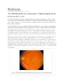
Webvision-EOG-Creel | The Electro-oculogram: Clinical Applications | The electrooculogram measures the potential that exists between the cornea and Bruch's membrane at the back of the eye. The potential produces a dipole field with the cornea approximately 5 millivolts positive compared to the back of the eye, in a normally illuminated room. Although the origin of th... | Electro-oculogram |
| 211 |
 |
Webvision-ERG-Creel | The Electroretinogram and Electro-oculogram: Clinical Applications | The global or full-field electroretinogram (ERG) is a mass electrical response of the retina to photic stimulation. The ERG is a test used worldwide to assess the status of the retina in eye diseases in human patients and in laboratory animals used as models of retinal disease. | Electroretinogram; Electro-oculogram |
| 212 |
 |
Webvision-mfERG-Creel | The Multifocal Electroretinogram: Clinical Applications | The most important development in ERGs is the multifocal ERG (mfERG). Erich Sutter adapted the mathematical sequences called binary m-sequences creating a program that can extract hundreds of focal ERGs from a single electrical signal. This system allows assessment of ERG activity in small areas of ... | Multifocal Electroretinogram |
| 213 |
 |
Figure-06 | The Normal Pupillary Light Reflex | The normal pupillary light reflex is initiated following exposure to light. After a brief latency, both the right (solid line) and left (broken line) pupil constrict, then undergo a small amount of redilation (escape), followed by oscillations (hippus) if the light is sustained. Hippus is not a path... | Reflex, Pupillary; Examination, Pupillary |
| 214 |
 |
OV_Tom_Oberg_Orbital_Exam | The Orbital Exam | Comprehensive demonstration of the entire orbital examination. | Orbital Examination |
| 215 |
 |
walsh_2000_c30 | The Wall-Eyed Potato Farmer | Young man presenting with apparent episodic neurologic evants that initially was thought to be multiple sclerosis, but as time went on, he had progressive changes in his neurologic exam and in his imaging findings. Brain biopsy revealed Gliomatosis cerebri. Anatomy: Brain Stem; Pons; Midbrain. Patho... | Gliomatosis Cerebri; Intracranial Tumors; Bilateral Internuclear Ophthalmoplegia |
| 216 |
 |
NOVEL_Moran_3a-15 | Third Nerve Palsy | Patient with third nerve palsy (no audio) | Third Nerve Palsy |
| 217 |
 |
1-5 | Third Nerve Palsy, Pupil Involving | Example of patient with third nerve palsy. Left eye shows pupilary involvement. Left eye doesn't immediately duct, but abducts well, with impaired superduction. Secondary and primary deviations are demonstrated. Anisocoria is more prominent when light is on, showing a parasympathetic defect to the p... | Pupil; Third Nerve Palsy; Third Nerve Dysfunction |
| 218 |
 |
Tilted_Discs_KBD.pdf | Tilted Discs | Short PowerPoint discussion of tilted discs with illustrations and images. | Tilted Disc |
| 219 |
 |
How2use | Tour of the Direct Ophthalmoscope | This clip describes the parts and operation of the ophthalmoscope as an ocular examination tool. Includes adjustment of aperture size and adjustment of lenses. | Direct Ophthalmoscope; Examination, Ocular |
| 220 |
 |
TheTour | Tour of the Fundus | This clip demonstrates the funduscopic examination technique. | Fundus; Examination, Ocular; Normal Optic Disc; AVP Macula; AVP Optic Nerve; Ophthalmoscopes |
| 221 |
 |
1-28 | Transillumination - Ciliary Body Neurofibromas | Example of transillumination on a patient with neurofibromatosis, but without Lisch nodules. Shows suspected neurofibromas in the ciliary body. | Transillumination; Examination, Ocular; Ciliary Body Neurofibromas1; Neurofibromatosis1 |
| 222 |
 |
1-29 | Transillumination - Lisch Nodules | Demonstration of transillumination of the Lisch nodules on a patient with neurofibromatosis. Shows how Lisch nodules that were not very visible in slit-lamp examination are better seen with transillumination, which may therefore be useful in detecting Lisch nodules earlier in children where they are... | Transillumination; Examination, Ocular; Lisch Nodules; Neurofibromatosis1 |
| 223 |
|
ocular_melanoma | Transillumination Ocular Melanoma | Video describing condition. | Ocular Melanoma |
| 224 |
 |
trigeminal_nerve_exam | Trigeminal Nerve Exam | Explanation of a trigeminal nerve exam. | Trigeminal Nerve |
| 225 |
 |
Tunnel_Vision_on_Tangent_Screen_Testing | Tunnel Vision on Tangent Screen Testing | Description of tunnel vision and tangent screen testing. | Tunnel Vision; Tangent Screen |