Best known for his world-renowned neuro-ophthalmology unit based at the University of California, San Francisco, William Hoyt, MD collected here more than 850 of his best images covering a wide range of disorders.
William F. Hoyt, MD, Professor Emeritus of Ophthalmology, Neurology and Neurosurgery, Department of Ophthalmology, University of California, San Francisco.
NOVEL: https://novel.utah.edu/
TO
| Title | Description | Type | ||
|---|---|---|---|---|
| 201 |
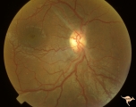 |
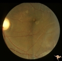
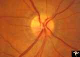
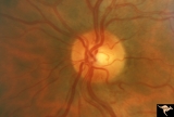
ID05b Post Papilledema Optic Atrophy from Pseudotumor Cerebri | Right eye, October 1999, Post papilledema optic atrophy from pseudotumor cerebri. Note optociliary veins in both discs. Gliosis and partial pallor following long standing papilledema and intracranial pressure. Anatomy: Optic disc. Pathology: Post papilledema atrophy and gliosis from long standing el... | Image |
| 202 |
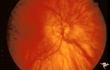 |
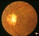
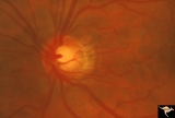
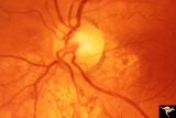
Venous Anomalies - Exit Anomalies | Choriovaginal vein. Inferior portion of disc at 6:00. Wide angle view of same patient as V_41. Anatomy: Optic disc. Pathology: Congenital anomaly of choroidal venous drainage. Disease/Diagnosis: Choriovaginal vein. Clinical: Asymptomatic. | Image |
| 203 |
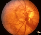 |
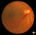
Venous Anomalies - Exit Anomalies | Choriovaginal vein. Inferior portion of disc at 5:00. Same patient as V_42. Anatomy: Optic disc. Pathology: Congenital anomaly of choroidal venous drainage. Disease/Diagnosis: Choriovaginal vein. Clinical: Asymptomatic. | Image |
| 204 |
 |
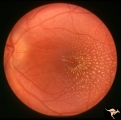
Venous Anomalies - Exit Anomalies | Anomalous exits of retinal veins at 5:00 . Anatomy: Optic disc. Pathology: Congenital anomaly, exit anomaly. Disease/Diagnosis: Exit anomaly, edge veins. Clinical: Asymptomatic. | Image |
| 205 |
 |
Diffuse Atrophy | Nerve fiber appearance about 6 weeks after indirect injury to optic nerve. Note near total absence of nerve fiber reflexes. Photo shows remaining streaks of inferior arcuate nerve fiber membranes dissolving into nothing. 1972. Anatomy: Optic disc. Pathology: Optic nerve injury. Disease/Diagnosis: Op... | Image |
| 206 |
 |
IA03 Atrophy with Optociliary Veins | 1974, left eye, perioptic nerve sheath meningioma, blind eye. Optociliary bypass veins in the nasal disc tissue. Anatomy: Optic disc. Pathology: Optociliary vein. DIsease/ Diagnosis: Perioptic nerve sheath meningioma. Clinical: Blind eye. | Image |
| 207 |
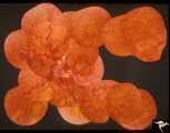 |
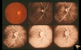
Vascular Disc Anomalies - Retinal Arteriovenous Malformations | Retinal arteriovenous malformations. Composite photograph. Notice there are at least two complete loops, nasally and infratemporally. Notice the signs of early involution centrally (white). Anatomy: Optic disc. Pathology: Retinal arteriovenous malformation. Disease/Diagnosis: Retinal arteriovenous m... | Image |
| 208 |
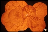 |
Vascular Disc Anomalies - Retinal Arteriovenous Malformations | Retinal arteriovenous malformations showing a complete arteriovenous loop nasally. 19 year old female. Composite photograph. Anatomy: Optic disc. Pathology: Retinal arteriovenous malformation. Disease/Diagnosis: Retinal arteriovenous malformation. Clinical: Asymptomatic. | Image |
| 209 |
 |
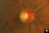
IB108 Post Ischemic (AION) Cupless Atrophy | March 1978, right eye, patient had inferior field defect, top half of optic nerve is atrophic, hemiatrophy, blood vessels are focally narrow, bottom half of disc is normal. Anatomy: Optic disc. Pathology: Post ischemic (AION) cupless atrophy. Disease/ Diagnosis: Post ischemic (AION) cupless atrophy.... | Image |
| 210 |
 |
Segmental Atrophy - Temporal | Segmental Atrophy - Temporal - Nutritional Amblyopia - Nerve fiber layer hemorrhage. Pair with IIA2_04b. 1970. Anatomy: Optic disc. Pathology: Optic atrophy. Disease/Diagnosis: Toxic optic atrophy from alcohol. Clinincal: Central visual loss. | Image |
| 211 |
 |
Segmental Atrophy - Temporal | Temporal - Nutritional Amblyopia - Nerve fiber layer hemorrhage. Pair with IIA2_04a. 1970. Anatomy: Optic disc. Pathology: Optic atrophy. Disease/Diagnosis: Toxic optic atrophy from alcohol. Clinical: Central visual loss. | Image |
| 212 |
 |
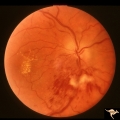
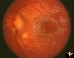
C201 Papillitis with Macular Star, Cat Scratch Disease | Proven Bartonella neuroretinitis. Notice the deposit of exudates of Henle's layer making an almost complete macular star. Anatomy: Optic disc; Retina. Pathology: Neuroretinitis; Axoplasmic stasis due to inflammation. Disease/ Diagnosis: Neuroretinitis due to Bartonella Henslae. Clinical: Visual blur... | Image |
| 213 |
 |
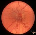
B110 Disc Swelling, Ischemic Papillopathies, AION | Pallid ischemic swelling with intraretinal exudates near the macula and a ""cotton wool"" infarct below the disc. 38 year old man. Diabetic. 20/60 vision. Altitudinal visual field defect. Anatomy: Optic disc. Pathology: Axoplasmic stasis due to ischemia. Disease/ Diagnosis: AION. Clinical: Diabetic ... | Image |
| 214 |
 |
B113 Disc Swelling, Ischemic Papillopathies, AION | 57 year old woman with AION. Anatomy: Optic disc. Pathology: Axoplasmic stasis due to ischemia. Disease/ Diagnosis: AION. Clinical: Visual loss. | Image |
| 215 |
 |
F2b10 Malignant Optic Nerve Glioma, Gross Pathologic Specimen | Pathologic specimen of optic nerve glioma shown in slide F2b_09. White material on top of swollen disc is myelin. Reference: Hoyt WF, Meshel LG, Lessell S, Schatz NJ, Suckling RD. Malignant optic glioma of adulthood. Brain. 1973;96(1):121-32. Anatomy: Optic disc. Pathology: Optic nerve glioma. Disea... | Image |
| 216 |
 |
Segmental Atrophy - Temporal | Segmental Atrophy - Temporal - Nutritional Amblyopia (alcohol) Discs show bilateral temporal pallor with hyperemia of the remaining disc tissue - Pair with IIA2_02b. 1971. Anatomy: Optic disc. Pathology: Optic atrophy. Disease/Diagnosis: Toxic optic atrophy from alcohol. Clinical: Central visual lo... | Image |
| 217 |
 |
Segmental Atrophy - Temporal | Segmental Atrophy - Temporal - Nutritional Amblyopia (alcohol) Discs show bilateral temporal pallor with hyperemia of the remaining disc tissue - Pair with IIA2_02a. 1971. Anatomy: Optic disc. Pathology: Optic atrophy. Disease/Diagnosis: Toxic optic atrophy from alcohol. Clinical: Central visual lo... | Image |
| 218 |
 |
Segmental Atrophy - Temporal | Segmental Atrophy - Temporal - Nutritional amblyopia (alcoholic). 1985. Left eye. Pair with IIA2_03a. Anatomy: Optic disc.. Pathology: Optic atrophy. Disease/Diagnosis: Toxic optic atrophy from alcohol. Clinical: Central visual loss. | Image |
| 219 |
 |
Segmental Atrophy - Hemianopic (Band) Atrophy from Eight Optic Tract Injury | Segmental Atrophy - Band atrophy from right optic tract injury. This eye has a nasal hemianopia. Its disc shows temporal pallor with an intact nasal nerve fiber layer. Pair with IIA2C_7b. Anatomy: Optic disc. Pathology: Right optic tract injury. Disease/Diagnosis: Homonymous hemioptic atrophy. Clini... | Image |
| 220 |
 |
Segmental Atrophy - Hemianopic (Band) Atrophy from Eight Optic Tract Injury | Segmental Atrophy - Band atrophy from right optic tract injury. Left eye. Has temporal hemianopia with band atrophy. Note loss of nasal nerve fiber layer. Four and a half months after injury from intracranial pressure catheter. Pair with IIA2C_7a. Anatomy: Optic disc. Pathology: Right optic tract in... | Image |
| 221 |
 |
Segmental Atrophy - Temporal | Segmental Atrophy - Temporal pallor - Nutritional amblyopia (alcoholic). 1985. Right eye. Pair with IIA2_03b. Anatomy: Optic disc. Pathology: Optic atrophy. Disease/Diagnosis: Toxic optic atrophy from alcohol. Clinical: Central visual loss. | Image |
| 222 |
 |
C207 Papillitis with Macular Star, Cat Scratch Disease | Proven Bartonella neuroretinitis. Anatomy: Optic disc; Retina. Pathology: Axoplasmic stasis due to inflammation. Disease/ Diagnosis: Bilateral Bartonella Henslae (Cat Scratch). Clinical: Visual blurring; Ocular disc edema with macular star (ODEMS). | Image |
| 223 |
 |
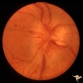
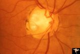
H33 Dysplasia with Hypoplasia (Elevated Dysplasia with Anomalous Vessels) | Left eye. Elevated dysplasia, hypoplasia. Pseudo papilledema. Woman. Congenital optociliary bypass at 7:00. Anatomy: Optic disc. Pathology: Dysplasia of the optic disc. Disease/ Diagnosis: Elevated dysplasia with hypoplasia. | Image |
| 224 |
 |
C02 Pits of the Optic Disc | Right eye. Three congenital optic pits on the temporal side. 8:00, 9:30, 10:30. Anatomy: Optic disc. | Image |
| 225 |
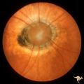 |
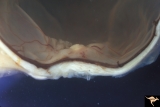
C11 Morning Glory Disc | "Morning Glory" disc. 11 year old girl. May not have a central retinal artery. Anatomy: Optic disc. | Image |