John A. Moran Eye Center Neuro-Ophthalmology Collection: A variety of lectures, videos and images relating to topics in Neuro-Ophthalmology created by faculty at the Moran Eye Center, University of Utah, in Salt Lake City.
NOVEL: https://novel.utah.edu/
TO
Filters: Collection: "ehsl_novel_jmec"
| Title | Description | Type | ||
|---|---|---|---|---|
| 201 |
 |
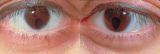
Aberrant Regeneration of the Seventh Nerve | Examples of patients with aberrant regeneration of the seventh nerve. First example is a patient with contractions around the mouth and dimpling, demonstrated with slow and rapid eye blinking. Second example shows contraction around nose with eye blink. | Image/MovingImage |
| 202 |
 |
Glaucoma: The Basics | Glaucoma is the most common optic neuropathy. Progressive cupping of the optic disc due to increased intraocular pressure together with visual field abnormalities and local disc susceptibility factors characterize this neuropathy. This PowerPoint lecture covers the basics of Glaucoma and includes ma... | Text |
| 203 |
 |
Optic Disc Pallor Pseudo and Real | Discussion of the causes of optic disc pallor. | Text |
| 204 |
 |
Testing the Visual Fields | Demonstration of various methods of testing visual fields, including counting fingers, motion, and color of several objects. | Image/MovingImage |
| 205 |
 |
Pulsating Exophthalmos | Example of a patient with neurofibromatosis with an absent sphenoid wing. Shows left eye pulsating back and forth with the pulse from front and side views. | Image/MovingImage |
| 206 |
 |
Facial Myokymia Unilateral | Example of patient with facial myokymia, a disorder of the seventh nerve, probably due to brain stem involvement. Patient has multiple sclerosis. Discussion of characteristics, such as continuous, undulating, contractions in the distribution of the seventh nerve, and a spreading of these movements t... | Image/MovingImage |
| 207 |
 |
Hemifacial Spasm | Example of patients with hemifacial spasm. First patient has a sequela of Bell's palsy, and is seen to have mainly clonic movements around the eye, with occasional tonic movements around the mouth. Second patient has a cerebellopontine angle epidurmoid tumor, and is seen to have movements around the... | Image/MovingImage |
| 208 |
 |
Bilateral Facial Myokymia | Example of a patient with a brain stem glioma. Shows bilateral facial myokymia. | Image/MovingImage |
| 209 |
 |
Pupillogram of a Healthy Young Subject | Pupillogram of a healthy young subject showing continuous pupillary oscillations of both pupils when light is sustained, indicated by the dark arrow at the top of the recording. Note that the oscillations of the pupils are synchronous and demonstrate variable amplitude and frequency. This pattern of... | Image |
| 210 |
 |
Relationship Between Age and Pupil Size | Relationship between age and pupil size, determined using an infrared flash photograph technique with subjects placed in darkness for 3 minutes. The numbers above the abscissa indicate the number of subjects tested in each age range. (Reprinted with permission of Loewenfeld IE: "Simple, central" ani... | Image |
| 211 |
 |
Normal Light Reflex without RAPD | This clip demonstrates the examination of the Relative Afferent Pupillary Defect (RAPD.) Demonstration of gauging the size of the pupil in light, testing light reflexes, swinging flashlight test for optic nerve abnormality. | Image/MovingImage |
| 212 |
 |
Enhanced Mydriasis in Response to Hydroxyamphetamine | Enhanced mydriasis in response to hydroxyamphetamine in a 77-year-old woman with a long-standing, preganglionic, right-sided Horner's syndrome that occurred following cervical neck dissection for thoracic outlet syndrome 30 years earlier. Miosis of the right pupil is apparent in room light (top). Th... | Image |
| 213 |
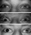 |
Left-sided Horner's Syndrome with an Acquired Preganglionic Localization | Left-sided Horner's syndrome in a 12-year-old girl with an acquired preganglionic localization based on clinical and pharmacologic testing. The cause remained undetermined after extensive radiologic investigations. Left-sided ptosis and miosis are evident in room light (top), and the degree of aniso... | Image |
| 214 |
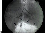 |
Bilateral Iris Colobomas (B) | Bilateral iris colobomas. B. Bilateral colobomatous defects of the inferonasal retina (black arrows) are also present, as shown in the right eye. | Image |
| 215 |
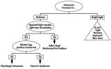 |
Flow Chart for Sorting Out Anisocoria - Bright Light and Darkness | Flow chart for sorting out anisocoria based initially on how it is influenced by bright light and darkness. | Image |
| 216 |
 |
Flow Chart for Sorting Out Anisocoria - Direct Light Reaction of the Pupil | Flow chart for sorting out anisocoria based initially on the integrity of the direct light reaction of the pupil. | Image |
| 217 |
 |
Assessment of an Afferent Pupillary Defect When Only One Iris is Functional | Assessment of an afferent pupillary defect when only one iris is functional. In this example, a right-sided parasellar tumor is compressing both the optic and oculomotor nerves, causing an optic neuropathy and a pupil-involving third crainial nerve palsy. The pupil on the affected side has both an a... | Image |
| 218 |
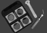 |
Hand-held Equipment Used to Measure a Relative Afferent Pupillary Defect | Hand-held equipment used to measure a relative afferent pupillary defect and to record pupil sizes. Four neutral density filters (0.3, 0.6, 0.9, 1.2 log units) are conveniently carried in a soft cloth carrying pouch. A bright light source (a Finhoff model illuminator is shown here) is ideal for stim... | Image |
| 219 |
 |
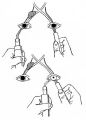
How to Measure the RAPD | This clip demonstrates the examination technique for measuring the Relative Afferent Pupillary Defect (RAPD). Demonstration of balancing an afferent papillary defect using filters in a patient with a resolving optic neuritis and an afferent papillary defect on the left. | Image/MovingImage |
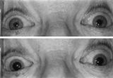
| 220 |
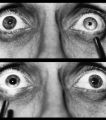 |
Right-sided Relative Afferent Pupillary Defect | Right-sided relative afferent pupillary defect in a man with optic nerve glioma. When the unaffected left eye is stimulated by light, both pupils constrict (top). When the light is then swung over to the affected right eye, both pupils dilate (bottom). This indicates that pupillomotor conduction thr... | Image |
| 221 |
 |
RAPD Present | This clip demonstrates the technique used to determine that Relative Afferent Pupillary Defect (RAPD) is present in a patient. | Image/MovingImage |
| 222 |
 |
Bilateral Iris Colobomas | Coloboma literally means a "gap"-and can be used to describe any fissure, hole, or gap in the eye. The term most often is used to refer to a congenital gap in the disc, retina, the choroid, and the iris. Colobomas occur because the embryonic fissure fails to fuse. Since the fissure closure begins in... | Image |
| 223 |
 |
Third Nerve Palsy, Pupil Involving | Example of patient with third nerve palsy. Left eye shows pupilary involvement. Left eye doesn't immediately duct, but abducts well, with impaired superduction. Secondary and primary deviations are demonstrated. Anisocoria is more prominent when light is on, showing a parasympathetic defect to the p... | Image/MovingImage |
| 224 |
 |
Aberrant Regeneration of the Lid | Patient with left third nerve palsy demonstrates anisocoria and mild vertical gaze limitation and aberrant movement of the left upper lid. Patient is instructed through all gaze positions. Left upper lid does not descend during downgaze but retracts instead. | Image/MovingImage |
| 225 |
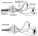 |
Pathophysiology of Signs Associated with a Tonic Pupil | Pathophysiology of signs associated with a tonic pupil. Normally, all parasympathetic fibers of the third cranial nerve synapse in the ciliary ganglion (top). Most postganglionic fibers innervate the ciliary muscle (dashed lines). After injury to the ciliary ganglion, the pupil becomes denervated an... | Image |