The Health Education Assets Library (HEAL) is a collection of over 22,000 freely available digital materials for health sciences education. The collection is now housed at the University of Utah J. Willard Marriott Digital Library.
TO
Filters: Collection: "ehsl_heal" Format: image
| Title | Description | Subject | Collection | ||
|---|---|---|---|---|---|
| 201 |
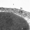 |
The air-blood barrier in the lung (rat) | Electron microscopy (high magnification). The alveolar space (1) and the capillary space (2) (with part of an erythrocyte, 3) are separated from each other respectively by cytoplasm of endothelial cell (5), the common basal lamina (6) with a lamina densa (6a) and the type I alveolar cell (4, pneumo... | Pneumocyte I; Alveolar cell type I | Poja Histology Collection - Respiratory System Subset |
| 202 |
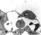 |
Brush cell in lung (rat) | Electron microscopy. In the proximal as well as in the terminal airways a special type of cell the so-called brush(-border) cell could be observed. In this picture this cell (X) is located in the alveolar space. At (*) the brush border, the dense cytoplasm contains many organelles. (C) is a capillar... | Brush cells | Poja Histology Collection - Respiratory System Subset |
| 203 |
 |
Elastin in lung arteriole (human, adult) | Immuno-electron microscopy (embedded in Lowicryl HM 20). The alveolar septa contain a muscular pulmonary arteriole (lumen = X) with unlabelled endothelial cells (1). The elastic membranes in the arteriolar walls consist of more or less continuous bands of amorphous elastic lumps (E) antibody-labeled... | Alveolar septum; Immuno-electron microscopy; Immuno-staining | Poja Histology Collection - Respiratory System Subset |
| 204 |
 |
Pseudoglandular - canalicular period of developing lung (human, fetus) | Stain: Azan. Location of a cartilagineous ring (1) between two cross-sectioned bronchi (2). Note in the bronchial epithelial cells the apical position of the epithelial nuclei and the light-stained basal part (↑) containing glycogen. The young hyaline cartilage presents solitary chondrocytes embed... | Lung development; Pseudoglandular period; Canalicular period; Mesenchyme | Poja Histology Collection - Respiratory System Subset |
| 205 |
 |
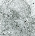
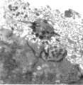
Alveolar macrophage in lung (rat) | Electron microscopy. A wandering alveolar macrophage (1) migrates through an alveolar pore from an alveolar space (A1) into another (A2). Note the dark lysosomal structures and abundant organelles in its cytoplasm. The thin lining type I alveolar cell is hardly discernable (thin arrows →). Interst... | Pneumocyte I; Alveolar macrophages | Poja Histology Collection - Respiratory System Subset |
| 206 |
 |
Epithelium of trachea (golden hamster) | Electron microscopy. Columnar ciliated cells (1) with cilia and organelle-rich apex; a goblet cell (2a) with accumulation of mucous secretion granules supranuclearly. Another goblet cell (2b) appears deprived; one basal cell (3) does not extend to the free surface. Light-grey aggregations of elastin... | Respiratory epithelium ; Pseudostratified epithelium; Basal cells | Poja Histology Collection - Respiratory System Subset |
| 207 |
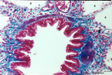 |
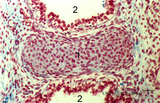
Bronchiolus in the lung (human, adult) | Stain: Azan. The lumen of the bronchiolus is lined with one layer of ciliated epithelium (1) and is folded due to the contraction of smooth muscle fibers (2) in the wall. Note the rich cellularity (3) in the stromal surrounding of the bronchiolus. Alveolar space (4). (5) pulmonary artery branches. | Ciliated epithelium; Bronchiolus | Poja Histology Collection - Respiratory System Subset |
| 208 |
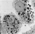 |
Alveolar macrophages in lung (human, adult) | Electron microscopy. Two macrophages in the alveolar space (N = nucleus). Note the heterogeneity of many lysosomal structures containing among others carbon particles and the abundancy of organelles. | Alveolar macrophages | Poja Histology Collection - Respiratory System Subset |
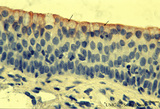
| 209 |
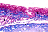 |
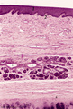
Section of trachea (human, adult, high magnification) | Stain: Azan. On top: the epithelium (1) is pseudostratified with cilia and goblet cells (white droplets) on a distinct basement membrane (↓, undulating thick deep-red line) followed by a small proper lamina (2). The submucosa (3) starts where the fibrous tissue (elastic fibers reinforced with coll... | Pseudostratified epithelium ; Tracheal glands; Excretory duct; Perichondrium | Poja Histology Collection - Respiratory System Subset |
| 210 |
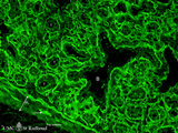 |
Laminin in the canalicular period of the lung (mouse, embryo) | Stain: Fluorescence microscopy with anti-laminin antibody. Laminin is a component of the basement membrane. Strong fluorescence visualizes the course of all basement membranes, especially around the bronchioles (B) and their branches in the developing lung tissue. In between the distal air spaces (t... | Bronchioli; Canalicular period | Poja Histology Collection - Respiratory System Subset |
| 211 |
 |
Multilamellar bodies of type II alveolar cell in the lung (mouse) | Electron microscopy. The cytoplasm of the type II alveolar cell (pneumocyte II) contains characteristic electron-dense multilamellar bodies (*) in different maturing stages. The lamellar bodies are responsible for the vacuolated appearance of these cells, and they give rise to surfactant (phospholip... | Pneumocyte II; Multilamellar bodies; Type II alveolar cell | Poja Histology Collection - Respiratory System Subset |
| 212 |
 |
Upper part of nasal septum (dog, higher magnification) | Stain: Hematoxylin and eosin. Stratified squamous epithelium (1) with small papillae (2). The lamina propria contains many small and large thin-walled blood vessels also known as the venous plexus (sinusoids) of Auerbach (3, swell body). Nasal glands (4) with draining ducts (5, larger diameter) and ... | Squamous stratified epithelium; Nasal vestibulum; Venous sinusoids; Venous plexus; Swell bodies | Poja Histology Collection - Respiratory System Subset |
| 213 |
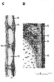 |
Scheme of trachea | C. Trachea (human, adult) magnification x 7.5 objective). D. Trachea (human, adult, detail of rectangle in C) magnification x 85 objective. (12) pseudostratified ciliated epithelium; (13) seromucous tracheal glands; (14) condensed layer of elastic fibers at the border of lamina propria and submu... | Pseudostratified epithelium ; Fibroelastic membrane; Adventitia | Poja Histology Collection - Respiratory System Subset |
| 214 |
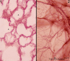 |
Elastic fibers in the lung (human) | Stain: Orcein. At the left (1) several alveoli (A) are depicted with a faint staining of cellular elements. The elastic fibers within the alveoli are stained reddish-purple. At the right (2) a high magnification of a tangential-sectioned alveolus shows the thicker bundles as well as very thin branch... | Elastic fibers | Poja Histology Collection - Respiratory System Subset |
| 215 |
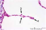 |
Alveolar septum in the lung (human, high magnification) | Stain: Azan. Two neighboring alveoli separated by a septum. (1) points to the tip containing bluish-pink elastin and collagen. The nucleus of a squamous alveolar cell (type I pneumocyte) is indicated by (2), and a free alveolar macrophage by (3). | Alveolar tip; Pneumocyte I | Poja Histology Collection - Respiratory System Subset |
| 216 |
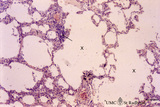 |
Progressing centrilobular lung emphysema (human, adult) | Stain: Hematoxylin and eosin. Emphysema is defined as enlargement of the air spaces (X) distal to the terminal bronchioles, with destruction of the alveolar walls. The remaining alveolar walls are thickened (1). There is an increased cellularity and locally signs of chronic inflammation (↓) are pr... | Poja Histology Collection - Respiratory System Subset | |
| 217 |
 |
Scheme of peripheral lung parenchym (human, adult) | (1) respiratory bronchiolus; (2) alveolar duct; (3) alveolar sac is a virtual sac formed by several alveoli, but continuous with the alveolar duct; (4) alveoli which end in small tips (↓) with elastin interwoven with collagen. | Alveolar duct | Poja Histology Collection - Respiratory System Subset |
| 218 |
 |
Keratin 7 staining in squamous metaplasia of the epithelium of a bronchiolus (human, adult) | Stain: anti-keratin 7 antibody (Pan-Ck 7) immunoperoxidase staining (with aminoethylcarbazole (AEC) substrate). A red-brown staining with AEC indicates a positive reaction for cytokeratin 7. Note that the top layer only is stained positively for keratin 7 (↓). Upon squamous metaplasia the bronchio... | Bronchiolus; Squamous metaplasia; Immunoperoxidase; Immuno-reaction | Poja Histology Collection - Respiratory System Subset |
| 219 |
 |
The alveolar tip in lung tissue (human, adult) | Electron microscopy. The lung tip is covered by a flattened alveolar cell type I (↓1) and a similar neighboring bulging one with nucleus (2). In the alveolar tip amorph elastin (5) appears more electron-dense than the collagen fibers (Col) and is interwoven with it. Small foci of calcifications a... | Alveolar tip; Elastoblast | Poja Histology Collection - Respiratory System Subset |
| 220 |
 |
Alveolar cell type II (pneumocyte II) in an alveolus (dog) | Electron microscopy. At (X) the alveolar space, the bulging alveolar cell type II shows the characteristic multilamellar bodies (*, cytosomes) that contain precusor material of surfactant. The lamellar bodies are responsible for the vacuolated appearance of these cells, and they give rise to surfact... | Pneumocyte II; Alveolar cell type II | Poja Histology Collection - Respiratory System Subset |
| 221 |
 |
Olfactory vesicle (bulb) in the nose (gerbil) | Electron microscopy. A distinct olfactory bulb (1, vesicle) with horizontally extended cross-sectioned cilia (*) and basal bodies (↑) is surrounded by numerous microvilli and free olfactory cilia. Close to the first bulb a second one is about to protrude above the surface of the supporting cells b... | Olfactory epithelium; Olfactory vesicle | Poja Histology Collection - Respiratory System Subset |
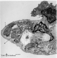
| 222 |
 |
Survey of bronchus and lung parenchym (human, adult) | Stain: Azan. (1) Lumen of small bronchus beside a lumen of pulmonary artery (2). Thin plates of hyaline cartilage (3) and connective tissue surrounding (1) and (2). Arrows (→) indicate lung alveoli with black-stained patches of carbon deposits. (4) Lung parenchym with alveoli. | Lung parenchym | Poja Histology Collection - Respiratory System Subset |
| 223 |
 |
Bronchiolus in the lung (human, adult) | Stain: Azan. The lumen is lined with one layer of ciliated epithelium (1) without goblet cells. Patches of smooth muscle fibers (2) are present. Notice small bronchial arteries (*). Arrows (↓) indicate sites of carbon accumulations between the alveoli (3). | Bronchiolus; Ciliated epithelium | Poja Histology Collection - Respiratory System Subset |
| 224 |
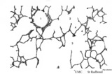
 |
Respiratory epithelium in the nasal vestibulum (rat) | Scanning electron microscopy. Note bushes of cilia (1) on the ciliary epithelium. In between areas of short microvilli-studded bulging or flattened goblet cells. Remnants of erythrocytes (2) are also found on top of ciliated cells. | Respiratory epithelium; Nasal vestibulum | Poja Histology Collection - Respiratory System Subset |
| 225 |
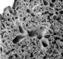 |
Survey of lung parenchym (gerbil) | Scanning electron microscopy. The photograph shows (1) bifurcation of several bronchi in the lung parenchym with alveoli (2). The long arrows indicate the route of alveolar ducts (4 ↕ length, and cross 4↑). Pulmonary vessels (3). | Lung parenchym; Pulmonary vessels | Poja Histology Collection - Respiratory System Subset |