The Health Education Assets Library (HEAL) is a collection of over 22,000 freely available digital materials for health sciences education. The collection is now housed at the University of Utah J. Willard Marriott Digital Library.
TO
| Title | Description | Subject | Collection | ||
|---|---|---|---|---|---|
| 201 |
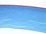 |
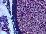
Epithelium - Cornea | The cornea of the eye is lined by simple squamous epithelium on its inner surface and stratified squamous epithelium on its outer surface. The stroma of the cornea consists of regularly arranged collagen fibers. UCLA Histology Collection. | simple squamous epithelium; stratified squamous epithelium | UCLA Histology |
| 202 |
 |
Cells / Organelles - Pituitary | Cells of the pituitary gland containing cytoplasmic secretory granules. The red-orange granules and the blue-purple granules contain different hormones. UCLA Histology Collection. | pars distalis; secretory granules | UCLA Histology |
| 203 |
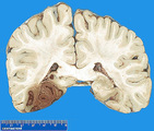 |
Recent hemorrhagic infarction | Recent hemorrhagic infarction | Knowledge Weavers Pathology | |
| 204 |
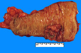 |
Adenocarcinoma, colon | Adenocarcinoma, colon | Knowledge Weavers Pathology | |
| 205 |
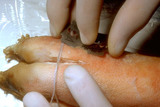 |
Shave technique | This demonstrates a shave technique on a pig's foot. Because there are no protruding lesions, I start by angling the blade at about 45 degrees from the skin surface, and while advancing the blade forward, I have a slight side to side sawing motion, and when I am about halfway through the target, I t... | Shave Biopsy | Knowledge Weavers Dermatology |
| 206 |
 |
Urticaria | This patient has developed rather severe urticaria, the cause of which was unknown. In urticaria, the skin swells and initially looks red and later can blanch as the amount of fluid increases within the skin. Internal organs can be involved in the process, and we are particularly concerned about the... | Knowledge Weavers Dermatology | |
| 207 |
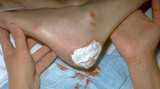 |
Silvadene | I placed Silvadene on the wound. Silvadene is an excellent antibacterial cream to apply to a wound after debridement. One must insure that an 1/8 inch to 1/4 inch coat is applied to help insure that the dressing doesn't absorb all the cream and allow the wound to subsequently dry out. | Knowledge Weavers Dermatology | |
| 208 |
 |
Ovary | A typical preantral follicle possessing a zona pellucida , multilayered granulosa or follicular cells and a stromal derived theca again embedded in stroma. Be sure you realize that the components within the zona pellucida are the nucleus of the ovum and its cytoplasm. UCLA Histology Collection. | Ovary; preantral follicle | UCLA Histology |
| 209 |
 |
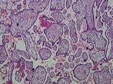
lymphoid tissue | This image shows stratified squamous epithelium overlying connective tissue. The parenchyma contains many lymphoid nodules. Crypts are present where epithelium invaginates into parenchyma. UCLA Histology Collection. | lymphoid nodule; lymphoid tissue; palatine tissue; palatine tonsil | UCLA Histology |
| 210 |
 |
Cells / Organelles - Liver | At a higher magnification, liver cell nuclei can be seen to have nucleoli. Note the heterochromatinheterochromatin lining the internal portion of the nuclear envelope. UCLA Histology Collection. | Cells and Organelles; Nucleus | UCLA Histology |
| 211 |
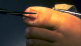 |
Ingrown nail | The nail plate is cut from the underlying nail bed with scissors. | Knowledge Weavers Dermatology | |
| 212 |
 |
Excision procedure | Demonstrates the same V. Summary) | Knowledge Weavers Dermatology | |
| 213 |
 |
Eye - Lens Fibers | Lens fibers elongating as they move posterior. UCLA Histology Collection. | UCLA Histology | |
| 214 |
 |
Peripheral Nervous System / Central Nervous System | The relationship between the spinal cord, the spinal roots, and the meninges is illustrated here. The surface of the spinal cord is covered by pia mater. Between the pia mater and web-like arachnoid mater is the subarachnoid space which contains CSF. The thick, collagenous dura mater is continuous w... | arachnoid mater; Central Nervous System; dura mater; meninges; Peripheral Nervous System; pia mater; spinal cord; spinal roots | UCLA Histology |
| 215 |
 |
Peripheral Nervous System | This cross sectioned peripheral nerve demonstrates the relatively thick myelin covering of axons, as well as the endoneurium which is located between the myelin sheaths. Also note the blue-stained collagen of the epineurium and a small blood vessel. UCLA Histology Collection. | axons; endoneurium; myelin; Peripheral Nervous System | UCLA Histology |
| 216 |
 |
Placenta | This image presents the basic organization of the placenta. Embedded within the maternal blood space are numerous fetal villi, some of them clearly tertiary villi , all lined by the syncytiotrophoblast . The only things missing in this image are maternal decidual cells and fetal/maternal fibrinoid. ... | Placenta | UCLA Histology |
| 217 |
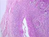 |
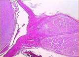
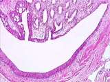
Epithelium - Lymphoid Tissue | Crypts of the palatine tonsils are lined by stratified squamous epithelium. The lymphoid tissue can be seen organized in nodules. A connective tissue capsule partially surrounds the tonsil. Note the skeletal muscle which is associated with the palatine tonsil. UCLA Histology Collection. | palatine tonsil | UCLA Histology |
| 218 |
 |
Kidney | The renal papilla is the site where terminal collecting ducts empty urine into the U-shaped minor calyx. The lumen of the minor calyx is lined by transitional epithelium. Strands of circular smooth muscle are found in the wall of the minor calyx. UCLA Histology Collection. | Kidney; minor calyx; renal papilla; terminal collecting ducts | UCLA Histology |
| 219 |
 |
Eye - Lens Epithelium | This enlargement of slide 236-2.5-2 reveals the lens epithelium in various phases: the anterior epithelium, area of proliferation, and area of elongation. The lens bow region consists of lens fiber nuclei in various stages of degeneration. UCLA Histology Collection. | UCLA Histology | |
| 220 |
 |
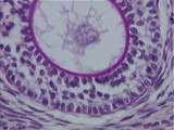
Thymus - Hassall's Corpuscle | This image shows an early and a late Hassall's corpuscle. Also visible are numerous epithelial reticular cells and even more numerous lymphocytes. UCLA Histology Collection. | epithelial reticular; Hassall's corpuscle | UCLA Histology |
| 221 |
 |
Adenocarcinoma, prostate | Adenocarcinoma, prostate | Knowledge Weavers Pathology | |
| 222 |
 |
Anterior pituitary | Anterior pituitary | Knowledge Weavers Pathology | |
| 223 |
 |
Circulatory System - Brachial Artery and Vein | This image shows the brachial artery (a muscular artery) at lower left, and the brachial vein at right. The staining of elastic fibers demonstrates the dark internal elastic membrane (IEM) and external elastic membrane (EEM), sandwiching the tunica media. The arterial intima appears to have sloughed... | brachial vein; tunica adventitia | UCLA Histology |
| 224 |
 |
Respiratory System | Bronchus-associated lymphoid tissue BALT consists of submucosal lymphoid tissue in intermediate and small airways. They are part of the diffuse mucosa-associated lymphoid tissue (MALT) also found in many other tissues. These structures are normal, but similar collections in lung alveolar tissue are ... | Lung; lymphoid tissue; Respiratory System | UCLA Histology |
| 225 |
 |
Placenta | This image presents all that you have to know about placental histology for this course. Fetal villi, some clearly tertiary villi , are completely surrounded by the maternal blood space. Lining everything in this image is the syncytiotrophoblast . The only other maternal component in this image is a... | Placenta | UCLA Histology |