Best known for his world-renowned neuro-ophthalmology unit based at the University of California, San Francisco, William Hoyt, MD collected here more than 850 of his best images covering a wide range of disorders.
William F. Hoyt, MD, Professor Emeritus of Ophthalmology, Neurology and Neurosurgery, Department of Ophthalmology, University of California, San Francisco.
NOVEL: https://novel.utah.edu/
TO
Filters: Collection: ehsl_novel_wfh
| Identifier | Title | Description | Subject | ||
|---|---|---|---|---|---|
| 176 |
 |
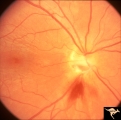
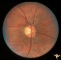
G1_02 | G102 Evulsion | Partial evulsion of the left optic nerve. Anatomy: Optic disc. Pathology: Optic nerve has been evulsed. Disease/ Diagnosis: Evulsion of the optic disc. | Disc Swelling; Traumatic Papillopathies; Evulsion; Optic Disc Evulsion |
| 177 |
 |
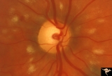
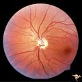
G1_03 | G103 Evulsion | Partial evulsion of the right optic nerve. Notice what is left of superior optic nerve. Anatomy: Optic disc. Pathology: Optic disc has been evulsed. Disease/ Diagnosis: Evulsion of the optic disc. | Disc Swelling; Traumatic Papillopathies; Evulsion; Optic Disc Evulsion |
| 178 |
 |
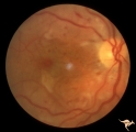
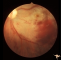
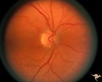
G2_04 | G204 Purtchers Traumatic Retinopathy | Right eye. Blind due to chest crush with broken ribs. 18 year old male. Anatomy: Optic disc. Pathology: Varied peripapillary ischemic retinopathy. Disease/ Diagnosis: Purtchers traumatic retinopathy. | Disc Swelling; Traumatic Papillopathies; Purtchers Traumatic Retinopathy |
| 179 |
 |
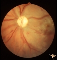
G2_05 | G205 Purtchers Traumatic Retinopathy | Right eye. Purtcher's retinopathy caused by chest crush from seat belt. Anatomy: Optic disc. Pathology: Varied peripapillary ischemic retinopathy. Disease/ Diagnosis: Purtchers traumatic retinopathy. | Disc Swelling; Traumatic Papillopathies; Purtchers Traumatic Retinopathy |
| 180 |
 |
G2_06 | G206 Purtchers Traumatic Retinopathy | Left eye. After auto accident in which the patient's chest was squeezed. Same eye as G2_07. Anatomy: Optic disc. Pathology: Varied peripapillary ischemic retinopathy. Disease/ Diagnosis: Purtchers traumatic retinopathy. | Disc Swelling; Traumatic Papillopathies; Purtchers Traumatic Retinopathy |
| 181 |
 |
G2_07 | G207 Purtchers Traumatic Retinopathy | Left eye. Large pre-retinal hemorrhage. Same eye as G2_06. Anatomy: Optic disc. Pathology: Varied peripapillary ischemic retinopathy. Disease/ Diagnosis: Purtchers traumatic retinopathy. | Disc Swelling; Traumatic Papillopathies; Purtchers Traumatic Retinopathy |
| 182 |
 |
G2_08 | G208 Traumatic AION | Traumatic vitreopapillary evulsion (traumatic AION). Traumatic AION from evulsion of the vitreopapillary adhesion. Leakage on disc surface where vitreous was adherent. Pair with G2_9b. Anatomy: Optic disc. Pathology: AION. Disease/ Diagnosis: Traumatic AION. | Disc Swelling; Traumatic Papillopathies; Vitreopapillary Evulsion; Traumatic AION |
| 183 |
 |
G2_09 | G209 Traumatic AION | Traumatic vitreopapillary evulsion (traumatic AION). Fluorescein angiogram shows petal shaped avascular zones on the surface of the disc. Pair with G2_8a. Anatomy: Optic disc. Pathology: AION. Disease/ Diagnosis: Traumatic AION. Imaging: Flourescein angiogram. | Disc Swelling; Traumatic Papillopathies; Vitreopapillary Evulsion; Traumatic AION |
| 184 |
 |
H_01 | H01 Panhypoplasia | Extreme hypoplasia. Very small disc. Peri-papillary halo (choroidal). Right eye. Note: normal vessels. Same patient as H_2. Anatomy: Optic disc. Pathology: Hypoplasia of the optic nerve. Disease/ Diagnosis: Hypoplasia. Clinical: Blind child. | Optic Disc; Congenital Anomalies; Severe Hypoplasias |
| 185 |
 |
H_02 | H02 Panhypoplasia | Extreme hypoplasia. Very small disc. Peri-papillary halo (choroidal). Left eye. Note: normal vessels. Same patient as H_1. Anatomy: Optic disc. Pathology: Hypoplasia of the optic nerve. Disease/ Diagnosis: Hypoplasia. Clinical: Blind child. | Optic Disc; Congenital Anomalies; Severe Hypoplasias |
| 186 |
 |
H_03 | H03 Panhypoplasia | Extreme hypoplasia. Note absence of retinal nerve fiber layer. Left eye. Girl. Same patient as H_4. Anatomy: Optic disc. Pathology: Hypoplasia of the optic nerve. Disease/ Diagnosis: Hypoplasia. Clinical: Left eye. Girl. | Optic Disc; Congenital Anomalies; Severe Hypoplasias |
| 187 |
 |
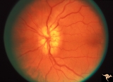
H_04 | H04 Panhypoplasia | Right eye. Normal eye. Girl. Same patient as H_3. Anatomy: Optic disc. Pathology: Hypoplasia of the optic nerve. Disease/ Diagnosis: Hypoplasia. | Optic Disc; Congenital Anomalies; Severe Hypoplasias |
| 188 |
 |
H_05 | H05 Panhypoplasia | Right eye. Distinctive septo-optic dysplasia.Hypoplasia of the optic nerve. Left eye normal. Amblyopic right eye. 24 year old woman. Anatomy: Optic disc. Pathology: Hypoplasia of the optic nerve. Disease/ Diagnosis: Hypoplasia. | Optic Disc; Congenital Anomalies; Severe Hypoplasias |
| 189 |
 |
H_06 | H06 Panhypoplasia | Bilateral hypoplasia. Top is Right eye - moderate. Bottom is Left eye - severe. Note venous tortuosity. Good example of double ring sign. De Morsier's syndrome.Septo-optic dysplasia. Same patient as H_7. Anatomy: Optic disc. Pathology: Hypoplasia of the optic nerve. Disease/ Diagnosis: Hypoplasia. ... | Optic Disc; Congenital Anomalies; Severe Hypoplasias |
| 190 |
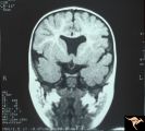 |
H_07 | H07 Panhypoplasia | MRI Scan, coronal view showing absence of septum pellucidum. Hypoplastic chiasm. De Morsier's syndrome. Same patient as H_6. Anatomy: Optic disc. Pathology: Hypoplasia of the optic nerve. Disease/ Diagnosis: Hypoplasia. Imaging: MRI scan. | Optic Disc; Congenital Anomalies; Severe Hypoplasias |
| 191 |
 |
H_08 | H08 Panhypoplasia | Severe hypoplasia. Right eye. Boy. Good example of double ring sign. Anatomy: Optic disc. Pathology: Hypoplasia of the optic nerve. Disease/ Diagnosis: Hypoplasia. | Optic Disc; Congenital Anomalies; Severe Hypoplasias |
| 192 |
 |
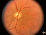
H_09 | H09 Panhypoplasia | Moderate hypoplasia. Man. Anatomy: Optic disc. Pathology: Hypoplasia of the optic nerve. Disease/ Diagnosis: Hypoplasia. | Optic Disc; Congenital Anomalies; Moderate Hypoplasias |
| 193 |
 |
H_10 | H10 Panhypoplasia | Cruzon's Disease. 26 year old man. Right eye. Mild hypoplasia. Son of patient in H_11 and H_12. Same patient in H_31. Father of patient in H_32. Anatomy: Optic disc. Pathology: Hypoplasia of the optic nerve. Disease/ Diagnosis: Hypoplasia. | Optic Disc; Congenital Anomalies; Mild Hypoplasias |
| 194 |
 |
H_101 | H101 Occipital Hemianoptic Hypoplasia | Right eye. Same patient as H_102. Anatomy: Optic disc. Pathology: Occipital hemianoptic hypoplasia. Disease/ Diagnosis: Congenital defect of the occipital lobe. | Optic Disc; Congenital Anomalies; Segmental Hypoplasia; Occipital Hemianoptic Hypoplasia |
| 195 |
 |
H_102 | H102 Occipital Hemianoptic Hypoplasia | Left eye. Trans-synaptic band atrophy. Left homonymous hemianopia from right occipital porencephaly. Loss of nasal nerve fibers. Same patient as H_101. Anatomy: Optic disc. Pathology: Occipital hemianoptic hypoplasia. Disease/ Diagnosis: Congenital defect of the occipital lobe. | Optic Disc; Congenital Anomalies; Segmental Hypoplasia; Occipital Hemianoptic Hypoplasia |
| 196 |
 |
H_103 | H103 Occipital Hemianoptic Hypoplasia | Right eye. Congenital right homonymous hemianopia. Absent nerve fiber layer in right eye. Same patient as H_104. Anatomy: Optic disc. Pathology: Occipital hemianoptic hypoplasia. Disease/ Diagnosis: Congenital defect of the occipital lobe. | Optic Disc; Congenital Anomalies; Segmental Hypoplasia; Occipital Hemianoptic Hypoplasia |
| 197 |
 |
H_104 | H104 Occipital Hemianoptic Hypoplasia | Left eye. Contrast with nasal nerve fiber in right eye, H_103. Anatomy: Optic disc. Pathology: Occipital hemianoptic hypoplasia. Disease/ Diagnosis: Congenital defect of the occipital lobe. | Optic Disc; Congenital Anomalies; Segmental Hypoplasia; Occipital Hemianoptic Hypoplasia |
| 198 |
 |
H_105 | H105 Occipital Hemianoptic Hypoplasia | Left congenital homonymous hemianopia. Right occipital AVM. Nasal nerve fiber layer loss in left eye. Compare with right eye. Same patient as H_106. Anatomy: Optic disc. Pathology: Occipital hemianoptic hypoplasia. DIsease/ Diagnosis: Congenital defect of the occipital lobe | Optic Disc; Congenital Anomalies; Segmental Hypoplasia; Occipital Hemianoptic Hypoplasia |
| 199 |
 |
H_106 | H106 Occipital Hemianoptic Hypoplasia | Same patient as H_105. Anatomy: Optic disc. Pathology: Occipital hemianoptic hypoplasia. Disease/ Diagnosis: Congenital defect of the occipital lobe. | Optic Disc; Congenital Anomalies; Segmental Hypoplasia; Occipital Hemianoptic Hypoplasia |
| 200 |
 |
H_11 | H11 Panhypoplasia | Cruzon's Disease. 47 year old woman. Right eye. Mild hypoplasia. Mother of patient in H_10 and H_31. Same patient as H_12. Grandmother of patient in H_32. Anatomy: Optic disc. Pathology: Hypoplasia of the optic nerve. Disease/ Diagnosis: Hypoplasia. | Optic Disc; Congenital Anomalies; Mild Hypoplasias |