Best known for his world-renowned neuro-ophthalmology unit based at the University of California, San Francisco, William Hoyt, MD collected here more than 850 of his best images covering a wide range of disorders.
William F. Hoyt, MD, Professor Emeritus of Ophthalmology, Neurology and Neurosurgery, Department of Ophthalmology, University of California, San Francisco.
NOVEL: https://novel.utah.edu/
TO
| Title | Description | Type | ||
|---|---|---|---|---|
| 176 |
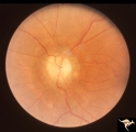 |
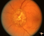
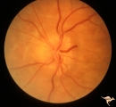
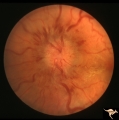
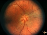
Chronic Papilledema with Pseudo Drusen | Left eye. Meningioma. Pseudo drusen from chronic papilledema. The patient's meningioma had blinded her left eye and caused chronic elevated intracranial pressure. Woman. Anatomy: Optic disc Pathology: Papilledema Disease/Diagnosis: Chronic papilledema with pseudo drusen | Image |
| 177 |
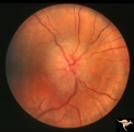 |
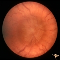
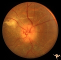
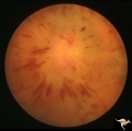
D101 Disc Edema with Systemic Lupus | Unilateral disc swelling with narrowed arterioles. No decrease in visual acuity or field. 19 year old woman. Patient died of cerebral lupus within two months. Optociliary veins dumping into disc edge at 4:00, 9:00, and 11:00. Anatomy: Optic disc. Pathology: Axoplasmic stasis due to vasculitis (Lupu... | Image |
| 178 |
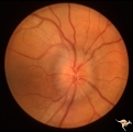 |
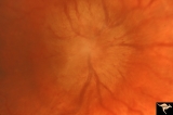
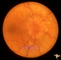
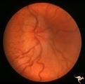
D102 Disc Edema with Systemic Lupus | 28 year old woman. Vision 20/20 but blind spot enlarged. Same patient as D1_03. Right eye. Anatomy: Optic disc. Pathology: Axoplasmic stasis due to vasculitis (Lupus). Disease/ Diagnosis: Lupus papillopathy. Clinical: Normal vision with enlarged blind spot on visual field. | Image |
| 179 |
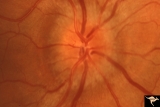 |
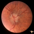
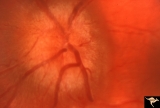
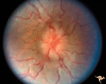
D103 Disc Edema with Systemic Lupus | 28 year old woman with systemic Lupus erythematosus. Vision 20/20 but blind spot enlarged. Same patient as D1_02. Magnified. Anatomy: Optic disc. Pathology: Axoplasmic stasis due to vasculitis (Lupus). Disease/ Diagnosis: Lupus papillopathy. Clinical: Normal vision with enlarged blind spot on visual... | Image |
| 180 |
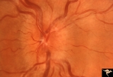 |
D104 Disc Edema with Systemic Lupus | Unilateral disc swelling and enlarged blind spot. Patient had episcleritis 4 weeks before this image was taken. 14 year old girl. Anatomy: Optic disc. Pathology: Axoplasmic stasis due to vasculitis (Lupus). Disease/ Diagnosis: Lupus papillitis. Clinical: No visual loss. History of episcleritis. Big ... | Image |
| 181 |
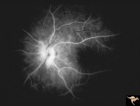 |
D106 Disc Edema with Systemic Lupus | Flourescein angiogram shows evidence of vascular papillopathy. (Lupus) Same patient as D1_05 an D1_07. Anatomy: Optic disc. Pathology: Axoplasmic stasis due to vasculitis (Lupus). Disease/ Diagnosis: Lupus papillopathy. | Image |
| 182 |
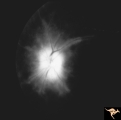 |
D107 Disc Edema with Systemic Lupus | Late stage Flourescein angiogram showing flourescein leakage on the disc and around the neighboring vessels. Note this amount of edema could not be appreciated in the colored fundus image D1_05. Same patient as D1_06 an D1_05. Anatomy: Optic disc. Pathology: Axoplasmic stasis due to vasculitis (Lupu... | Image |
| 183 |
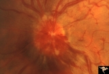 |
E01 Disc Swelling with Central Vein Occlusion | Left eye. Central retinal vein occlusion with disc swelling. Anatomyt: Optic disc; Retina. Pathology: Vasculitis. Disease/ Diagnosis: Disc swelling due to retinal vasculitis. | Image |
| 184 |
 |
Early Papilledema due to Brain Tumor - Resolving | Left eye. Same eye as P_34a. One month post op, papilledema resolving. Boy. Anatomy: Optic disc. Pathology: Papilledema. Disease/Diagnosis: Papilledema from posterior fossa hemangioblastoma. | Image |
| 185 |
 |
F104 Esthesio Neuroblastoma | Esthesioneuroblastoma. Tumor cells infiltrating the optic disc. 20/200 vision. Anatomy: Optic disc. Pathology: Esthesioneuroblastoma. Disease/ Diagnosis: Esthesioneuroblastoma. | Image |
| 186 |
 |
F105 Histiocytosis Infiltrate of Disc | Histiocytosis infiltrate of right disc with simultaneous infiltration of the hypothalamus with skin lesions on eye lids and chest. Same patient as F1_06. Anatomy: Optic disc. Pathology: Histiocytosis infiltrate. Disease/ Diagnosis: Histiocytosis infiltrate. Clinical: Patient presented with skin lesi... | Image |
| 187 |
 |
F106 Histiocytosis Infiltrate of Disc | More fully developed and chronic histiocytosis infiltrate of right disc with simultaneous infiltration of the hypothalamus with skin lesions on eye lids and chest. Same patient as F1_05, one year later. Anatomy: Optic disc. Pathology: Histiocytosis infiltrate of disc. Disease/ Diagnosis: Histiocytos... | Image |
| 188 |
 |
F109 T-Cell Leukemia Infiltrate | T-Cell leukemia infiltrate. 14 year old boy with T-Cell leukemia infiltrating the disc. Anatomy: Optic disc. Pathology: T-Cell leukemia. Disease/ Diagnosis: Neoplastic (metastatic) papillopathy | Image |
| 189 |
 |
F201 Optic Nerve Sheath Meningioma | Right eye. Woman with ophthalmoplegia proptosis for 14 years. Visual field reduced due to optic nerve sheath meningioma. Notice large optociliary vessel temporally. Anatomy: Optic disc. Pathology: Chronic optic disc swelling caused by optic nerve sheath meningioma. Disease/ Diagnosis: Chronic optic ... | Image |
| 190 |
 |
F203 Optic Nerve Sheath Meningioma | Optic nerve sheath meningioma. Note optociliary vessels on the disc. The disc is partially atrophic and blurred by previous edema. The cause of the choroidal scar was not determined. Anatomy: Optic disc. Pathology: Chronic optic disc swelling caused by optic nerve sheath meningioma. DIsease/ Diagnos... | Image |
| 191 |
 |
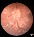
F207 Disc Swelling due to Metastatic Breast Cancer | Unilateral disc swelling with retinal folds due to metastatic breast cancer. Apparent enophthalmus. Anatomy: Optic disc. Pathology: Metastatic breast cancer. Disease/ Diagnosis: Neoplastic papillopathy. | Image |
| 192 |
 |
F2b01 Optic Nerve Glioma | Left eye. Woman with optic nerve glioma. Anatomy: Optic disc. Pathology: Optic nerve swelling secondary to retrobulbar optic glioma. Disease/ Diagnosis: Optic nerve glioma. | Image |
| 193 |
 |
F2b02 Progressive Optic Disc Swelling with Optic Glioma | Progressive optic disc swelling with optic glioma. Left eye. Woman with optic disc swelling. April 1969. Same patient as F2b_03 and F2b_04. Anatomy: Optic disc. Pathology: Optic nerve swelling secondary to retrobulbar optic glioma. Disease/ Diagnosis: Optic nerve glioma. | Image |
| 194 |
 |
F2b03 Progressive Optic Disc Swelling with Optic Glioma | Progressive optic disc swelling with optic glioma. Left eye. Woman with optic disc swelling. Edema is becoming pale. May 1969. Same patient as F2b_02 and F2b_04. Anatomy: Optic disc. Pathology: Optic nerve swelling secondary to retrobulbar optic glioma. Disease/ Diagnosis: Optic nerve glioma. | Image |
| 195 |
 |
F2b04 Progressive Optic Disc Swelling with Optic Glioma | Progressive optic disc swelling with optic glioma. Left eye. Woman with optic disc swelling. Entire disc obscured by overlying edema and hemorrhage. Blind in 3 months. This series illustrates a progressive infarction of the optic disc adjacent to an optic disc glioma. June 1969. Same patient as F2b_... | Image |
| 196 |
 |
F2b05 Optic Disc Swelling from Optic Glioma | Optic disc swelling from optic glioma. Patient had Neurofibromatosis (NF1). Left eye. 7 year old girl. 20/100 acuity. Glioma of the left optic nerve. Anatomy: Optic disc. Pathology: Optic nerve glioma. Disease/ Diagnosis: Optic nerve swelling secondary to retrobulbar optic glioma | Image |
| 197 |
 |
F2b06 Optic Disc Swelling from Optic Glioma | Right eye. Optic glioma with disc swelling. Anatomy: Optic disc. Pathology: Optic nerve glioma. Disease/ Diagnosis: Optic nerve swelling secondary to retrobulbar optic glioma. | Image |
| 198 |
 |
F2b07 Optic Disc Swelling from Optic Glioma | 6 year old with neurofibromatosis (NF1). Right eye went blind. Light perception. Optic canal enlargement due to glioma. Notice optociliary vessels. Same patient as F2b_8. Anatomy: Optic disc. Pathology: Optic nerve glioma. Disease/ Diagnosis: Optic nerve swelling secondary to retrobulbar optic gliom... | Image |
| 199 |
 |
F2b08 Optic Disc Swelling from Optic Glioma | Left eye. Optic nerve glioma. Disc swelling without visual loss. Same patient as F2b_7. Anatomy: Optic disc. Pathology: Optic nerve glioma. Disease/ Diagnosis: Optic nerve swelling secondary to retrobulbar optic glioma. | Image |
| 200 |
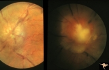 |
F2b09 Optic Disc Swelling from Malignant Optic Nerve Glioma | Malignant optic nerve glioma of adulthood with blindness and optic disc edema. Right image shows white material extruded from the swollen optic disc. This material is myelin being squeezed into the eye from the nerve infarction. Autopsy specimen of this eye shown in F2b_10. Reference: Hoyt WF, Meshe... | Image |