Best known for his world-renowned neuro-ophthalmology unit based at the University of California, San Francisco, William Hoyt, MD collected here more than 850 of his best images covering a wide range of disorders.
William F. Hoyt, MD, Professor Emeritus of Ophthalmology, Neurology and Neurosurgery, Department of Ophthalmology, University of California, San Francisco.
NOVEL: https://novel.utah.edu/
TO
Filters: Collection: "ehsl_novel_wfh"
| Title | Description | Type | ||
|---|---|---|---|---|
| 176 |
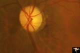 |
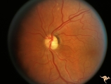
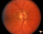
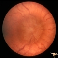
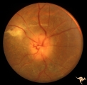
Diffuse Atrophy | Primary optic atrophy from optic nerve compression by aneurysm. Note narrowing of retinal arterioles. Pair with IIA1_8. Anatomy: Optic disc. Pathology: Optic atrophy. Disease/Diagnosis: Optic atrophy due to giant aneurysm. Clinical: Blindness. | Image |
| 177 |
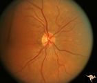 |
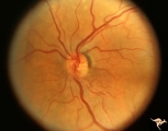
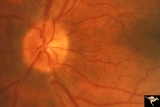
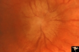
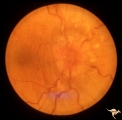
Diffuse Atrophy - Evolution of Optic Disc Palor After Optic Nerve Transection | Evolution of optic disc pallor after optic nerve transection. Normal Right eye. Photo taken December 9, 1978. Anatomy: Optic disc. Pathology: Total retrograde optic atrophy. Disease/Diagnosis: Transection of the optic nerve. Clinical: Blindness. | Image |
| 178 |
 |
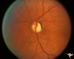
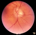
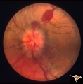
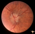
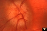
Diffuse Atrophy - Evolution of Optic Disc Palor After Optic Nerve Transection | Injury on December 8, 1978. Evolution of optic disc pallor after optic nerve transection. Woman having rhinoplasty suffered optic nerve transection. Left eye. Photo taken January 11, 1979 - 33 days post accident. Note superior and inferior arcuate nerve fiber bundles are thinned. Optic disc shows s... | Image |
| 179 |
 |
Diffuse Atrophy - Evolution of Optic Disc Palor After Optic Nerve Transection | Injury on December 8, 1978. Evolution of optic disc pallor after optic nerve transection. Woman having rhinoplasty suffered optic nerve transection. Left eye. Photo taken February 14, 1979 - 65 days post accident. Optic disc is completely pale. All evidence of retinal nerve fiber layer is gone. Anat... | Image |
| 180 |
 |
Diffuse Atrophy - Evolution of Optic Disc Palor After Optic Nerve Transection | Injury on December 8, 1978. Evolution of optic disc pallor after optic nerve transection. Woman having rhinoplasty suffered optic nerve transection. Left eye. Photo taken January 18, 1979 - 40 days post accident. Retinal nerve fiber layer appears thinner and disc is paler. Anatomy: Optic disc. Patho... | Image |
| 181 |
 |
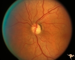
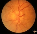
Diffuse Atrophy - Evolution of Optic Disc Palor After Optic Nerve Transection | Injury on December 8, 1978. Evolution of optic disc pallor after optic nerve transection. Woman having rhinoplasty suffered optic nerve transection. One day after nerve transection. Note dilated veins. Left eye. Photo taken December 9, 1978. Anatomy: Optic disc. Pathology: Total retrograde optic atr... | Image |
| 182 |
 |
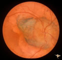
Drusen Plus Papilledema | PP37a: right swollen disc on top of drusen with narrowing of the arterioles;PP37 b: left visible drusen and papilledema with sub-retinal hemorrhage temporally. Patient had frontal glioblastoma. Anatomy: Optic disc. Pathology: Drusen of the optic disc. Disease/Diagnosis: Drusen of the optic disc. Cli... | Image |
| 183 |
 |
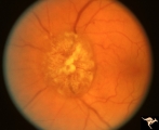
Drusen with Sub-retinal Neovascular Net | Buried drusen with sub-retinal neovascular net. There may be retinoschisis as well. Anatomy: Optic disc. Pathology: Drusen plus neovascularization at the border of the optic disc. Disease/Diagnosis: Drusen of the optic disc. Clinical: Patient has very large blind spot and impaired central vision. | Image |
| 184 |
 |
E01 Disc Swelling with Central Vein Occlusion | Left eye. Central retinal vein occlusion with disc swelling. Anatomyt: Optic disc; Retina. Pathology: Vasculitis. Disease/ Diagnosis: Disc swelling due to retinal vasculitis. | Image |
| 185 |
 |
E03 Disc Swelling with Central Retinal Vein Occlusion | 36 year old woman with visual obscurations of right eye. Early CRVO, papillophlebitis. Steroid responsive. Anatomy: Optic disc; Retina. Pathology: Central retinal vein occlusion. Disease/ Diagnosis: Disc swelling due to central retinal vein occlusion. Clinical: Decreased vision in right eye. Acuity ... | Image |
| 186 |
 |
E08 Disc Swelling with Central Vein Occlusion | Pituitary adenoma with right chronic CRVO with optociliary bypass vessels. Anatomy: Optic disc; Retina. Pathology: Central retinal vein occlusion. Disease/ Diagnosis: Disc swelling due to central retinal vein occlusion. | Image |
| 187 |
 |
E09 Disc Swelling with Central Vein Occlusion | Chronic disc swelling due to CRVO. Anatomy: Optic disc; Retina. Pathology: Central retinal vein occlusion. Disease/ Diagnosis: Disc swelling due to central retinal vein occlusion. | Image |
| 188 |
 |
E11 Disc Swelling with Central Vein Occlusion | Old retinal vein occlusion with optociliary bypass vessel at 3:00. Right eye. Anatomy: Optic disc; Retina. Pathology: Central retinal vein occlusion. Disease/ Diagnosis: Disc swelling due to central retinal vein occlusion. | Image |
| 189 |
 |
E12 Disc Swelling with Central Vein Occlusion | 2nd attack of papillophlebitis. There is an optociliary bypass vessel at 4:00. Anatomy: Optic disc; Retina. Pathology: Central retinal vein occlusion. Disease/ Diagnosis: Disc swelling due to central retinal vein occlusion. | Image |
| 190 |
 |
Early Papilledema due to Brain Tumor - Resolving | Left eye. Same eye as P_34a. One month post op, papilledema resolving. Boy. Anatomy: Optic disc. Pathology: Papilledema. Disease/Diagnosis: Papilledema from posterior fossa hemangioblastoma. | Image |
| 191 |
 |
Early Papilledema due to Tumor | Left eye. Asymmetric Papilledema with posterior fossa hemangioblastoma. Left - moderate papilledema. Blurring of disc. Young man. Anatomy: Optic disc. Pathology: Papilledema. Disease/Diagnosis: Papilledema from posterior fossa hemangioblastoma. | Image |
| 192 |
 |
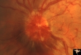
F104 Esthesio Neuroblastoma | Esthesioneuroblastoma. Tumor cells infiltrating the optic disc. 20/200 vision. Anatomy: Optic disc. Pathology: Esthesioneuroblastoma. Disease/ Diagnosis: Esthesioneuroblastoma. | Image |
| 193 |
 |
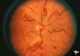
F105 Histiocytosis Infiltrate of Disc | Histiocytosis infiltrate of right disc with simultaneous infiltration of the hypothalamus with skin lesions on eye lids and chest. Same patient as F1_06. Anatomy: Optic disc. Pathology: Histiocytosis infiltrate. Disease/ Diagnosis: Histiocytosis infiltrate. Clinical: Patient presented with skin lesi... | Image |
| 194 |
 |
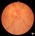
F106 Histiocytosis Infiltrate of Disc | More fully developed and chronic histiocytosis infiltrate of right disc with simultaneous infiltration of the hypothalamus with skin lesions on eye lids and chest. Same patient as F1_05, one year later. Anatomy: Optic disc. Pathology: Histiocytosis infiltrate of disc. Disease/ Diagnosis: Histiocytos... | Image |
| 195 |
 |
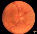
F109 T-Cell Leukemia Infiltrate | T-Cell leukemia infiltrate. 14 year old boy with T-Cell leukemia infiltrating the disc. Anatomy: Optic disc. Pathology: T-Cell leukemia. Disease/ Diagnosis: Neoplastic (metastatic) papillopathy | Image |
| 196 |
 |
F201 Optic Nerve Sheath Meningioma | Right eye. Woman with ophthalmoplegia proptosis for 14 years. Visual field reduced due to optic nerve sheath meningioma. Notice large optociliary vessel temporally. Anatomy: Optic disc. Pathology: Chronic optic disc swelling caused by optic nerve sheath meningioma. Disease/ Diagnosis: Chronic optic ... | Image |
| 197 |
 |
F203 Optic Nerve Sheath Meningioma | Optic nerve sheath meningioma. Note optociliary vessels on the disc. The disc is partially atrophic and blurred by previous edema. The cause of the choroidal scar was not determined. Anatomy: Optic disc. Pathology: Chronic optic disc swelling caused by optic nerve sheath meningioma. DIsease/ Diagnos... | Image |
| 198 |
 |
F207 Disc Swelling due to Metastatic Breast Cancer | Unilateral disc swelling with retinal folds due to metastatic breast cancer. Apparent enophthalmus. Anatomy: Optic disc. Pathology: Metastatic breast cancer. Disease/ Diagnosis: Neoplastic papillopathy. | Image |
| 199 |
 |
F2b01 Optic Nerve Glioma | Left eye. Woman with optic nerve glioma. Anatomy: Optic disc. Pathology: Optic nerve swelling secondary to retrobulbar optic glioma. Disease/ Diagnosis: Optic nerve glioma. | Image |
| 200 |
 |
F2b02 Progressive Optic Disc Swelling with Optic Glioma | Progressive optic disc swelling with optic glioma. Left eye. Woman with optic disc swelling. April 1969. Same patient as F2b_03 and F2b_04. Anatomy: Optic disc. Pathology: Optic nerve swelling secondary to retrobulbar optic glioma. Disease/ Diagnosis: Optic nerve glioma. | Image |