The Health Education Assets Library (HEAL) is a collection of over 22,000 freely available digital materials for health sciences education. The collection is now housed at the University of Utah J. Willard Marriott Digital Library.
TO
| Title | Description | Subject | Collection | ||
|---|---|---|---|---|---|
| 176 |
 |
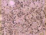
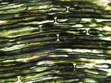
Tongue (lingual gland, human) | Stain: Azan. The posterior lingual gland is composed of areas with pure serous acini neighbouring pure mucous acini between the tongue muscles (at the left few striated fibers). | oral cavity; lingual gland | Poja Histology Collection - Oral Cavity Subset |
| 177 |
 |
Tongue (ventral part, human) | Stain: Hematoxylin and eosin. Non-keratinized squamous epithelium, followed by a small lamina propria and the intrinsic striated lingual muscles. | oral cavity; lingual muscles | Poja Histology Collection - Oral Cavity Subset |
| 178 |
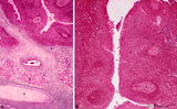 |
Tubal tonsil (human) | Stain: Azan. The tubal tonsil consists of a collection of lymphoid nodules near the auditory tube opening and forms part of the Waldeyers ring of defense in the nasopharyngeal cavity. This tonsil has fewer crypts (1), and the surface is covered by one to more layered ciliated epithelium (2). The la... | tubal tonsil; nasopharynx | Poja Histology Collection - Lymphatic Tissues and Organs Subset |
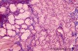
| 179 |
 |
Villi of complete hydatidiform mole (human) | (A) Macroscopy aggregation (1) of abnormal villi of hydatidiform mole intermingled with blood clots (2). (B) After dissection of a complete mole grape-like aggregations of dilated chorionic villi are presented as numerous liquid-filled vesicles (1) varying in diameters (from few mm up to 1 cm) surr... | placenta; trophoblast; hydatiform mole | Poja Histology Collection - Placenta |
| 180 |
 |
Trophoblast cell in lung alveolar interstitium (human, early midpregnancy) | Stain: Hematoxylin-eosin. It is well known that free circulating trophoblast cells can be found migrated into lung parenchyma during normal pregnancy. In this sample a large trophoblast cell (→) is localised within the normal alveolar interstitium. Alveolar phagocytes (*) are present in the alve... | placenta; trophoblast; lung | Poja Histology Collection - Placenta |
| 181 |
 |
Uvula (soft palate, human) | Stain: Azan. Detail of the uvula at the nasal side shows the border between the non-keratinized stratified squamous epithelium (right, oral side) and pseudostratified columnar epithelium (left, nasal side). The lamina propria is composed of dense connective tissue. At the bottom part of a blood-fil... | oral cavity; lining mucosa | Poja Histology Collection - Oral Cavity Subset |
| 182 |
 |
Uvula (soft palate, human; low magnification) | Stain: Azan. At the right (oral side, non-keratinized stratified squamous epithelium); at the left (nasal side, same epithelium). In the submucosa mainly mucous uvular glands are localized. At the bottom, striated muscle of the uvular muscle. The uvular tip is richly vascularized. | oral cavity | Poja Histology Collection - Oral Cavity Subset |
| 183 |
 |
Uvula (soft palate, human) | Stain: Azan. Detail of the uvula at the oral side; non-keratinized stratified squamous epithelium. Note richly vascularized lamina propria. In the submucosa mainly mucous uvular glands with draining ducts. | oral cavity; lining mucosa; mucous glands | Poja Histology Collection - Oral Cavity Subset |
| 184 |
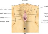 |
Vulva (Labeled) | External genitalia. Female. | Mons Pubis; Glans; External Urethral Orifice; Perineal Body; Opening of Vagina; Vestibule of Vagina; Labium Minus; Labium Majus | Royal College of Surgeons in Ireland Illustrations |
| 185 |
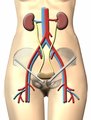 |
Urinary System | Urinary system. Kidneys. Ureter. Bladder. | Common Iliac Veins; Common Iliac Arteries | Royal College of Surgeons in Ireland Illustrations |
| 186 |
 |
Eye - Rods and Cones | Rod and cone photoreceptors have their cell bodies in the outer nuclear layer. Between these cell bodies and the retinal pigment epithelium are the photoreceptor inner segments (where proteins are synthesized) and the outer segments (the light sensitive portion). The inner segments of cones are cone... | UCLA Histology | |
| 187 |
 |
Uterus | This relatively brief phase of the endometrial cycle consists of shrinkage of the endometrium and intraendometrial hemorrhage and fragmentation. The myometrium is located to the left. A higher magnification of the ischaemic endometrium is shown in image 226-10-1. UCLA Histology Collection. | ischaemic endometrium; Uterus | UCLA Histology |
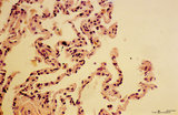
| 188 |
 |
Kidney | In this section through the renal papilla, identify the terminal collecting tubules (lined by columnar epithelium). Also note a straight segment of the loop of Henle, many thin loops of Henle, and cross-sections of the vasa recta. UCLA Histology Collection. | Kidney; renal papilla; terminal collecting tubules | UCLA Histology |
| 189 |
 |
Breast | This active, lactating breast contains a large number of lobules of tightly packed alveoli. The connective tissue of the interlobular elements is highly compressed. Note the lactating glands, the excretory duct and some fat. UCLA Histology Collection. | Breast; lactating breast | UCLA Histology |
| 190 |
 |
Suturing | After placing four throws to create a square knot, the suture is then cut just above the knot. | Knowledge Weavers Dermatology | |
| 191 |
 |
Bone - Haversian System | Haversian systems can be seen well in this image of dried compact bone. Haversian or central canals and concentrically arranged osteocytes are also visible. UCLA Histology Collection. | UCLA Histology | |
| 192 |
 |
Suturing | The suture is then tightened by crossing the non-dominant (left) hand over the dominant (right) hand. | Knowledge Weavers Dermatology | |
| 193 |
 |
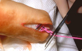
Suturing | This demonstrates tightening of the double loop along the long axis of the wound using suture. | Knowledge Weavers Dermatology | |
| 194 |
 |
Skin | Healing skin (rat). See lymphocyte infiltration. UCLA Histology Collection. | Skin | UCLA Histology |
| 195 |
 |
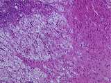
Somatotropinoma | Somatotropinoma | Knowledge Weavers Pathology | |
| 196 |
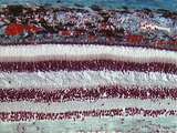 |
Eye - Retina | The retina consists of the retinal pigment epithelium and neurosensory retina. The neurosensory retina includes multiple layers of neurons and some glia. The outer nuclear layer contains the cell bodies of rod and cone photoreceptors. The inner nuclear layer contains the cell bodies of multiple neur... | UCLA Histology | |
| 197 |
 |
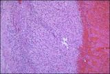
Ovary | This tissue looks like a dying corpus luteum, but that is a rare event. This is an example of a structure that we cannot identify unless we see a zona pellucida and/or a glassy membrane. If you had a section along the plane labelled A you would not be able to differentiate between a section through ... | Ovary | UCLA Histology |
| 198 |
 |
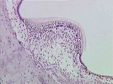
Corpus luteum cysts exhibit a convoluted lining with luteinized granulosa and theca cells. | Corpus luteum of the ovary, medium power. The granulosa cells have undergone proliferation and alteration to lutein cells that produce progesterone. The lutein cells are large and polyhedral and the cytoplasm is foamy and eosinophilic. | Knowledge Weavers Human Reproduction | |
| 199 |
 |
Ear | At this higher magnification, one can differentiate the hair & supporting cells, the gelatinous cupula, and the dark cells which mark the transition of the membranous labyrinth into the crista ampullaris. Bone is also evident. Movement of the endolymph against the cupula causes bending of hair cell ... | Crista Ampullaris; Ear | UCLA Histology |
| 200 |
 |
Peripheral Nervous System | This slide of myelinated nerves is stained with osmium to demonstrate the myelin sheaths. The region where two myelin sheaths covering the same axon contact one another is the Node of Ranvier. The region of myelin sheath between these nodes is known as the internode. UCLA Histology Collection. | myelin sheaths; osmium; Peripheral Nervous System | UCLA Histology |