A collection of videos relating to the diagnosis and treatment of eye movement disorders. This collection includes many demonstrations of examination techniques.
Dan Gold, D.O., Associate Professor of Neurology, Ophthalmology, Neurosurgery, Otolaryngology - Head & Neck Surgery, Emergency Medicine, and Medicine, The Johns Hopkins School of Medicine.
A collection of videos relating to the diagnosis and treatment of eye movement disorders.
NOVEL: https://novel.utah.edu/
TO
| Title | Description | Type | ||
|---|---|---|---|---|
| 176 |
 |
Pendular Nystagmus and Ocular Motor Signs in MS | 𝗢𝗿𝗶𝗴𝗶𝗻𝗮𝗹 𝗗𝗲𝘀𝗰𝗿𝗶𝗽𝘁𝗶𝗼𝗻: This is a 30-year-old man with a 15 year history of multiple sclerosis. For the last 12 months, he experienced horizontal oscillopsia. On examination, there were ocular motor abnormalities including gaze-evoked nystagmus,... | Image/MovingImage |
| 177 |
 |
Pendular Nystagmus and Vision Loss | Three patients are presented here, each with poor vision (counting fingers or worse) related to retinitis pigmentosa in one patient (Usher's syndrome) and optic neuropathy in two patients, each of whom developed pendular nystagmus after vision loss developed. Visually mediated movements normally pre... | Image/MovingImage |
| 178 |
 |
Pendular, Gaze-Evoked and Abducting Nystagmus in MS | This is a 40-year-old woman with a history of multiple sclerosis who presented for oscillopsia. On examination, she had bilateral internuclear ophthalmoplegia (INO-adduction lag OU and abducting nystagmus OU), with a corresponding exotropia that increased in right and left gaze. She also had horiz... | Image/MovingImage |
| 179 |
 |
Penlight Cover Test (Partial Removal of Fixation) | Penlight cover test (partial removal of fixation): during in-person clinical encounters, the maneuvers below are best tested with complete (or near complete) removal of fixation (e.g., Frenzel or video Frenzel goggles). Removal of fixation is more challenging during virtual evaluations but can be ap... | Image/MovingImage |
| 180 |
 |
Periodic Alternating Nystagmus and Central Head-Shaking Nystagmus from Nodulus Injury | 𝗢𝗿𝗶𝗴𝗶𝗻𝗮𝗹 𝗗𝗲𝘀𝗰𝗿𝗶𝗽𝘁𝗶𝗼𝗻: This is a 35-year-old man who suffered a gunshot wound to his cerebellum. When he regained consciousness days later, he experienced oscillopsia due to periodic alternating nystagmus (PAN). He was started on baclofen 10 mg... | Image/MovingImage |
| 181 |
 |
Periodic Alternating Nystagmus and Perverted Head-shaking Nystagmus in Cerebellar Degeneration | 𝗢𝗿𝗶𝗴𝗶𝗻𝗮𝗹 𝗗𝗲𝘀𝗰𝗿𝗶𝗽𝘁𝗶𝗼𝗻: This is a 60-yo-woman with several years of worsening imbalance, diplopia (hers was actually unrelated to cerebellar pathology [although she did have an esotropia greater at distance that was cerebellar in origin] and due... | Image/MovingImage |
| 182 |
 |
Periodic Alternating Nystagmus Due to a Chiari Malformation | This patient first experienced oscillopsia 12 months prior to this video. Three months after the onset of symptoms, she was seen by neuro-ophthalmology and found to have a spontaneous, unidirectional left-beating nystagmus (that did not reverse) in addition to saccadic smooth pursuit. Oscillopsia wo... | Image/MovingImage |
| 183 |
 |
Periodic Alternating Nystagmus Due to Nodulus Stroke | This is a 70-year-old woman who experienced the acute onset of vertigo and imbalance. MRI demonstrated a diffusion-weighted imaging hyperintensity involving the nodulus (with corresponding ADC hypointensity) consistent with an acute stroke. On examination several weeks after the stroke, periodic alt... | Image/MovingImage |
| 184 |
 |
Periodic Alternating Nystagmus Due to Spinocerebellar Ataxia Type 6 | 𝗢𝗿𝗶𝗴𝗶𝗻𝗮𝗹 𝗗𝗲𝘀𝗰𝗿𝗶𝗽𝘁𝗶𝗼𝗻: This 50-yo-man complained of imbalance for several years and more recently oscillopsia. On examination, there was saccadic pursuit and VOR suppression in addition to gaze-evoked nystagmus with rebound, raising suspicion f... | Image/MovingImage |
| 185 |
 |
Peripheral (Vestibular) and Central (Gaze-Evoked) Patterns of Nystagmus in a Single Patient | A 55-year-old man experienced episodic vertigo and was diagnosed with Meniere's disease affecting the left ear (based on audiograms and his clinical course) about 1 year prior to presentation. About 6 months prior to presentation, intratympanic (IT) gentamicin was injected into the left ear, at whic... | Image/MovingImage |
| 186 |
 |
Physiologic End Point Nystagmus | This is a normal subject with end point nystagmus in lateral gaze. Features that favor physiologic (normal) end point nystagmus (EPN) rather than pathologic gaze-evoked nystagmus include: only present in far lateral gaze (at close to 100% of the normal range of ocular movements); resolves when the v... | Image/MovingImage |
| 187 |
 |
Pinched Nose Valsalva | Valsalva (closed glottis or pinched nose): instruct the patient to take a deep breath and ‘bear down' (closed glottis) or take a deep breath and ‘try to pop their ears' (pinched nose). Assess for nystagmus. In superior canal dehiscence, pressure changes may be transmitted to the superior canal, ... | Image/MovingImage |
| 188 |
 |
Pontine Hemorrhage Causing Oculopalatal Tremor and Multiple Cranial Neuropathies | This is a 45-yo-woman who had a dorsal pontine cavernoma that bled 2 years prior to this video. Symptoms included diplopia and oscillopsia. On examination, she had left>right facial palsies (upper and lower face from involvement of the nucleus/fascicle - i.e., lower motor neuron palsies) and sixth n... | Image/MovingImage |
| 189 |
 |
Positional Downbeat Nystagmus Mimicking Anterior Canal BPPV | Although positional downbeat nystagmus (pDBN) can indicate the rare anterior canal variant of benign paroxysmal positional vertigo, central mimics are common causes of pDBN. pDBN may be seen in multiple system atrophy (MSA), or seen with posterior fossa lesions, with a common example being a stroke ... | Image/MovingImage |
| 190 |
|
Positional Nystagmus During an Attack of Vestibular Migraine | 𝗢𝗿𝗶𝗴𝗶𝗻𝗮𝗹 𝗗𝗲𝘀𝗰𝗿𝗶𝗽𝘁𝗶𝗼𝗻: A 50-year-old woman presented to clinic after experiencing multiple episodes of hours-long vertigo attacks that were associated with headache, photophobia and phonophobia. She had a history of motion sickness and migraine... | Image/MovingImage |
| 191 |
 |
Post-infectious Ocular Flutter and Myoclonus Syndrome | 𝗢𝗿𝗶𝗴𝗶𝗻𝗮𝗹 𝗗𝗲𝘀𝗰𝗿𝗶𝗽𝘁𝗶𝗼𝗻: This is a 35-yo-woman presenting with oscillopsia following a viral illness. She described being easily startled, with "shakiness" of the head/neck and body. She had myoclonus and ocular flutter, with the latter evident w... | Image/MovingImage |
| 192 |
 |
Posterior Canal - BPPV: Epley and Semont Maneuvers | 𝗢𝗿𝗶𝗴𝗶𝗻𝗮𝗹 𝗗𝗲𝘀𝗰𝗿𝗶𝗽𝘁𝗶𝗼𝗻: Epley/canalith repositioning maneuver (CRP) To treat right posterior canal (PC)-BPPV (each position maintained for at least 30 seconds or until nystagmus and/or vertigo cease): • First the patient is placed in the long-... | Image/MovingImage |
| 193 |
 |
Posterior Canal BPPV Pre- and Post-Epley Maneuver | 𝗢𝗿𝗶𝗴𝗶𝗻𝗮𝗹 𝗗𝗲𝘀𝗰𝗿𝗶𝗽𝘁𝗶𝗼𝗻: This is a patient with typical right posterior canal benign paroxysmal positional vertigo (BPPV), which was provoked by the Dix-Hallpike maneuver. When the patient was moved into the right Dix-Hallpike maneuver, after a b... | Image/MovingImage |
| 194 |
 |
Posterior Canal BPPV Treated with Semont Maneuver | This is a patient with left posterior canal (PC) benign paroxysmal positional vertigo (BPPV), and upbeat-torsional (towards the left ear) nystagmus was provoked by left Dix-Hallpike maneuver and left side-lying maneuver. This video demonstrates treatment of her left PC BPPV with the Semont maneuver.... | Image/MovingImage |
| 195 |
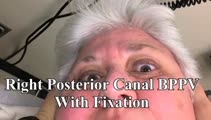 |
Posterior Canal BPPV with Fixation and with Fixation Removed | This is a 60-yo-woman with positional vertigo. In the right Dix-Hallpike position with fixation removed, there was clear upbeat-torsional nystagmus (towards the lowermost right ear) which led to the diagnosis of right posterior canal BPPV. In right Dix-Hallpike with fixation there was mainly torsion... | Image/MovingImage |
| 196 |
 |
Pressure Testing for Superior Canal Dehiscence Syndrome | 𝗢𝗿𝗶𝗴𝗶𝗻𝗮𝗹 𝗗𝗲𝘀𝗰𝗿𝗶𝗽𝘁𝗶𝗼𝗻: Superior semicircular canal dehiscence syndrome (SCDS) is caused by a third mobile window in the inner ear. This allows for transmission of sound or pressure to the superior canal. Tragal compression and/or glottic and ... | Image/MovingImage |
| 197 |
 |
Prolonged Lid Twitch in Myasthenia Gravis | This 50-yo-woman with ocular MG demonstrated a spontaneous and particularly prolonged eyelid twitch. | Image/MovingImage |
| 198 |
 |
Provocative Maneuvers (Removal of Fixation, Vibration, Head-Shaking) to Accentuate Peripheral Vestibular Nystagmus) | 𝗢𝗿𝗶𝗴𝗶𝗻𝗮𝗹 𝗗𝗲𝘀𝗰𝗿𝗶𝗽𝘁𝗶𝗼𝗻: With an acute destructive process like vestibular neuritis that causes significant unilateral vestibular loss, spontaneous nystagmus is always present. However, over days to months, spontaneous nystagmus should resolve co... | Image/MovingImage |
| 199 |
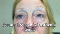 |
Pseudo-INOs in Myasthenia Gravis | This is a 55-yo-woman with an intermittent exotropia who had normal adduction OU, but clear lag of adducting saccades OD>OS with rapid horizontal saccades. This was much more apparent after repeat testing (ie, it was fatigable), and she wound up having ocular MG. | Image/MovingImage |
| 200 |
 |
Pseudo-Spontaneous Nystagmus and Bow and Lean Test in Horizontal Canal BPPV | 𝗢𝗿𝗶𝗴𝗶𝗻𝗮𝗹 𝗗𝗲𝘀𝗰𝗿𝗶𝗽𝘁𝗶𝗼𝗻:This is a 70-year-old woman presenting to the Emergency Department with positional vertigo that was determined to be due to the apogeotropic variant of right horizontal canal (HC) benign paroxysmal positional vertigo (BPPV... | Image/MovingImage |