The Health Education Assets Library (HEAL) is a collection of over 22,000 freely available digital materials for health sciences education. The collection is now housed at the University of Utah J. Willard Marriott Digital Library.
TO
Filters: Collection: ehsl_heal
| Title | Description | Subject | Collection | ||
|---|---|---|---|---|---|
| 151 |
 |
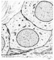
Epithelial lining of respiratory bronchiolus in the lung (rat) | Electron microscopy (low magnification). The epithelial lining of the respiratory bronchiolus contains ciliated cells (1) and Clara cells (2) with electron-dense secretory granules. At (4) interstitium with elastin, collagen and myofibroblasts; (5) shows a small pulmonary arteriole. At arrow (→) a... | Respiratory bronchiolus; Ciliated epithelium; Clara cells; Secretory granules; Pneumocytes; Alveolar cells; Interstitium | Poja Histology Collection - Respiratory System Subset |
| 152 |
 |
Epithelial lining of terminal bronchiolus (golden hamster) | Electron microscopy (low magnification). Two major epithelial cell types line the bronchiolus: (1) the ciliated cells with many organelles, and (2) the non-ciliated cells or Clara cells well provided with organelles, especially agranular endoplasmic reticulum and depending on their activity, numerou... | Terminal bronchiolus; Ciliated epithelium ; Clara cells; Neuro-endocrine cells; Pneumocytes; Alveolar cell types; Interstitium | Poja Histology Collection - Respiratory System Subset |
| 153 |
 |
Epithelial lining of trachea and bronchiolus (mammals) | Left side trachea: Columnar ciliated cells (1) with up to 200 motile cilia with an organelle-rich apex and in the middle a goblet cell (2) with accumulation of mucus secretion granules in the apex. Note that all epithelial cells (i.e., junctions) contact the basal lamina. Four basal cells (3) do not... | Bronchiolus; Pseudostratified epithelium; Ciliated epithelium; Clara cells; Basal cells; Neuro-endocrine cells | Poja Histology Collection - Respiratory System Subset |
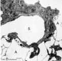
| 154 |
 |
Epithelial tooth bud in tooth development - tooth germ, human, embryo | Stain: hematoxylin. At the top stratified ectoderm. The proliferating basal cell layers are palisade-arranged with a light cytoplasm close to the basement membrane (right side). Below the basement membrane subepithelially an accumulation of inductive neural crest-derived mesenchymal cells is locally... | oral cavity; tooth bud; tooth development | Poja Histology Collection - Oral Cavity Subset |
| 155 |
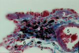 |
Epithelium of respiratory bronchiolus (detail, human, high magnification) | Stain: Azan. An irregular lining of low columnar-cuboidal cells (1) and thin alveolar epithelium (2) are present. Note cuboidal Clara cells (↓). At (3) small patches of smooth muscles and at (4) macrophages with phagocytized carbon particles (black dots). Small pulmonary artery (5), lymph capillar... | Respiratory bronchiolus; Cuboidal epithelium; Alveolar epithelium; Lymph capillary | Poja Histology Collection - Respiratory System Subset |
| 156 |
 |
Epithelium of trachea (golden hamster) | Electron microscopy. Columnar ciliated cells (1) with cilia and organelle-rich apex; a goblet cell (2a) with accumulation of mucous secretion granules supranuclearly. Another goblet cell (2b) appears deprived; one basal cell (3) does not extend to the free surface. Light-grey aggregations of elastin... | Respiratory epithelium ; Pseudostratified epithelium; Basal cells | Poja Histology Collection - Respiratory System Subset |
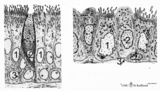
| 157 |
 |
Epithelium of trachea (mammals, common respiratory epithelium) | Scheme electron microscopy. Columnar ciliated cells (1) with up to 200 motile cilia with an organelle-rich apex and in the middle a goblet cell (2) with accumulation of mucus secretion granules in the apex. Note that all epithelial cells (i.e., junctions) contact the basal lamina. Four basal cells (... | Respiratory epithelium ; Pseudostratified epithelium; Basal cells | Poja Histology Collection - Respiratory System Subset |
| 158 |
 |
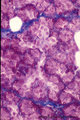
Erythroblast and neutrophilic myelocytes in bone marrow smear (human) | Stain: May-Grnwald-Giemsa (MGG). (1) an older basophilic erythroblast with slightly condensed nuclear chromatin. (2) two neutrophilic myelocytes with azurophilic primary granules. | Poja Histology Collection - Blood & Bone Marrow Subset | |
| 159 |
 |
Erythroblastic island (bone marrow, rabbit) | Electron microscopy. In the bone marrow close associations of developing red blood cells with reticular cells are required during erythropoiesis. Different stages of erythroblasts (2, 3) are exposed in close vicinity of a reticular cell (1). (4) Reticulocyte. The centrally localized phagocytic retic... | Poja Histology Collection - Blood & Bone Marrow Subset | |
| 160 |
 |
Erythroblastic island in bone marrow | Scheme electron microscopy. Different stages of maturing erythroblasts (2) are exposed in this scheme. The centrally localized phagocytic reticular cell (1) has many long cytoplasmic extensions that form a network with similar cells within the bone marrow. Its nucleus is irregular. The cytoplasm has... | Poja Histology Collection - Blood & Bone Marrow Subset | |
| 161 |
 |
Erythroblasts (bone marrow, rabbit) | Electron microscopy. (1) shows basophilic erythroblasts or early normoblasts with clumped heterochromatin in the nucleus and numerous polysomes. (1m) points to a mitotic figure of a basophilic erythroblast. A late polychromatic erythroblast or intermediate normoblast (2) marks a maturing stage in wh... | Poja Histology Collection - Blood & Bone Marrow Subset | |
| 162 |
 |
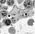
Erythrocytes in peripheral blood smear (human) | Stain: May-Grnwald-Giemsa (MGG). Normal erythrocytes in a blood smear of peripheral blood. The mature human erythrocytes do not have nuclei. Their normal diameter is about 7.5 μm. Macrocytes have a diameter >9 μm, while microcytes are smaller than 6 μm. The thickness of an erythrocyte in the cent... | Poja Histology Collection - Blood & Bone Marrow Subset | |
| 163 |
 |
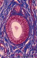
Erythron in bone marrow smear (human) | Stain: May-Grnwald-Giemsa (MGG). The erythron or erythroblastic island consists of a large reticulum cell (1) surrounded by erythroblastic cell types (2) at varoius stages of differentiation. The nucleus of the reticulum cell (histiocyte) usually contains a noticeable nucleolus. At distance, some my... | Poja Histology Collection - Blood & Bone Marrow Subset | |
| 164 |
 |
Erythron in bone marrow smear (human) | Stain: May-Grnwald-Giemsa (MGG). In the center of the slide an erythron that consists of a reticular cell (1) surrounded by erythroblasts (2) at various maturational stages. | Poja Histology Collection - Blood & Bone Marrow Subset | |
| 165 |
 |
Erythron or erythropoietic island in bone marrow smear (human) | Stain: May-Grnwald-Giemsa. The picture shows a reticular cell (1) surrounded by almost exclusively erythroblasts at different stages of development and maturation (2). The cells marked with (*) are myeloid cell types. The reticular cell has phagocytized nuclear debris of expulsed nuclei (→). The r... | Poja Histology Collection - Blood & Bone Marrow Subset | |
| 166 |
 |
Exocrine gland (salivary gland) | Scheme electronmicroscopy. Part of an acinus of mucous cells with different amount of mucous secretion. Supranuclearly the Golgi areas with maturing mucous secretion granules, basolateral the endoplasmic reticulum. At the top the lumen. The left cell shows an accumulation of the secretory droplets, ... | oral cavity; mucous gland | Poja Histology Collection - Oral Cavity Subset |
| 167 |
 |
Exocrine gland (salivary gland) | Scheme electronmicroscopy. Part of an acinus of serous cells, basolaterally a well developed rough endoplasmic reticulum and apically secretion granules with different maturity towards the small lumen. On top stages of early formed secretion granule (left) and more matured ones (right). Note small j... | oral cavity; serous gland | Poja Histology Collection - Oral Cavity Subset |
| 168 |
 |
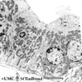
Exocrine gland - intercalated duct - submandibular gland, rat | Electronmicroscopy. The dark cells form an intercalated duct from the left lower corner to the right upper corner, and end in the acinus with lightly stained cells (serous). | oral cavity; intercalated duct; serous gland | Poja Histology Collection - Oral Cavity Subset |
| 169 |
 |
Exocrine gland - intercalated duct - salivary glands, pancreas | Scheme electronmicroscopy. Part of an intercalated duct, the low cuboidal epithelial cells contain sparsely organelles and some desmosomal structures. Between the lining cells and the basal lamina is squeezed part of a filament-rich myoepithelial cell. Lumen side top left quadrant. | oral cavity; intercalated duct | Poja Histology Collection - Oral Cavity Subset |
| 170 |
 |
Exocrine gland - intercalated duct - submandibular gland, human | Stain: Azan. Branching intercalated duct draining several serous acini, the lining duct cells are lighter stained due to the content of organelles. Serous cells are darker stained (many organelles) with round nuclei. Note few fat cells, the septa are blue stained. | oral cavity; intercalated duct; serous gland | Poja Histology Collection - Oral Cavity Subset |
| 171 |
 |
Exocrine gland - interlobular duct - submandibular gland, human | Stain: Azan. An interlobular duct with partly columnar as well as pseudostratified columnar epithelium. Note the dense connective tissue of the interlobular septum with small blood vessels. | oral cavity; seromucous glands | Poja Histology Collection - Oral Cavity Subset |
| 172 |
 |
Exocrine gland - parotid gland, rat | Electronmicroscopy. Part of a striated duct with basally membrane infoldings and numerous mitochondria perpendicularly orientated to the basal membrane. Upper side is luminal side. Apically junctional complexes as well as mitochondria and many vesicles are present. | oral cavity; serous gland | Poja Histology Collection - Oral Cavity Subset |
| 173 |
 |
Exocrine gland - striated (intralobular) duct - salivary glands, pancreas | Scheme electronmicroscopy. Part of a striated duct with basally membrane infoldings and mitochondria accumulations in the tall columnar cells. Small junctional complexes as well as apically mitochondria and numerous vesicles are present. | oral cavity; striated duct | Poja Histology Collection - Oral Cavity Subset |
| 174 |
 |
Exocrine gland - striated (intralobular) ducts - parotid gland, human | Stain: Azan. Cross-sections of striated ducts in upper part between serous acini. Note at the right bottom part of (lighter stained) lining cells of an intercalated duct. Connective tissues between the structures are blue stained. | oral cavity; striated duct | Poja Histology Collection - Oral Cavity Subset |
| 175 |
 |
Exocrine gland - submandibular gland, gerbil | Electronmicroscopy. Part of a serous acinus of this mixed gland. Note characteristic electron-dense secretion granules, the mature ones are larger. At the lower bottom is shown part of a lining cell of the intercalated duct. | oral cavity; serous gland | Poja Histology Collection - Oral Cavity Subset |