Best known for his world-renowned neuro-ophthalmology unit based at the University of California, San Francisco, William Hoyt, MD collected here more than 850 of his best images covering a wide range of disorders.
William F. Hoyt, MD, Professor Emeritus of Ophthalmology, Neurology and Neurosurgery, Department of Ophthalmology, University of California, San Francisco.
NOVEL: https://novel.utah.edu/
TO
Filters: Collection: "ehsl_novel_wfh"
| Title | Description | Type | ||
|---|---|---|---|---|
| 151 |
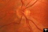 |
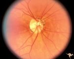
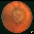
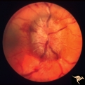
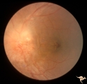
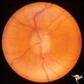
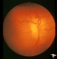
H34 Segmental Hypoplasia, Retinal-Congenital Toxo | Left eye. Temporal sector hypoplasia from congenital retinal toxoplasmosis. Note the sector shaped nerve fiber loss between 2:00 and 4:00. Same patient as H_35. Anatomy: Optic disc; retina. Pathology: Hypoplasia secondary to retinal lesion. Disease/ Diagnosis: Segmental optic disc hypoplasia. | Image |
| 152 |
 |
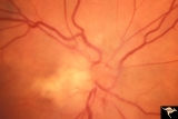
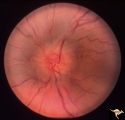
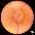
H36 Segmental Hypoplasia, Retinal, Congenital Toxo | Left eye. Optic disc hypoplasia from congenital nasal retinal toxoplasma lesion. Chorioretinal scar. Anatomy: Optic disc, retina. Pathology: Hypoplasia secondary to retinal lesion. Disease/ Diagnosis: Segmental optic disc hypoplasia. | Image |
| 153 |
 |
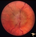
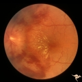
P55b Chronic Papilledema, Late Stage | Left eye. Optic atrophy secondary to chronic papilledema, late stage with retinal choroidal bypass vein. Opticocilliary shunt. Glioblastoma, chemotherapy patient. Anatomy: Optic disc. Pathology: Papilledema. Disease/ Diagnosis: Chronic papilledema with atrophy. | Image |
| 154 |
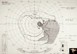 |
H51 Segmental Hypoplasia, Retinal-Nasal Hypoplasia | Visual field showing flag-like temporal field defect in patient shown in H_50. Anatomy: Optic disc. Pathology: Nasal segmental disc hypoplasia. Disease/ Diagnosis: Cogenital anomaly. | Image |
| 155 |
 |
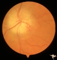
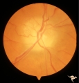
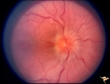
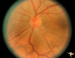
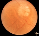
H50 Segmental Hypoplasia, Retinal-Nasal Hypoplasia | Right eye. Nasal hypoplasia with nasal pit. Same patient as H_51. Anatomy: Optic disc. Pathology: Nasal segmental disc hypoplasia. Disease/ Diagnosis: Congential anomaly. | Image |
| 156 |
 |
P55a Chronic Papilledema, Late Stage | Right eye. Optic atrophy secondary to chronic papilledema. Glioblastoma. Chemotherapy patient. Anatomy: Optic disc. Pathology: Papilledema. Disease/ Diagnosis: Chronic papilledema with atrophy. | Image |
| 157 |
 |
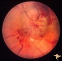
F101 Optic Disc Lymphosarcoma | Optic disc lymphosarcoma. This disc has been infiltrated by neoplastic cells. Anatomy: Optic disc. Pathology: Lymphosarcoma. Disease/ Diagnosis: Lymphosarcoma. | Image |
| 158 |
 |
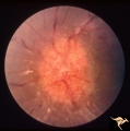
Bilateral Chronic Papilledema | Left eye. Frisen's stage 5. Patient with long standing aqueductal stenosis. Bilateral Chronic Papilledema. Man. Anatomy: Optic disc. Pathology: Papilledema. Disease/Diagnosis: Papilledema from aqueductal stenosis. | Image |
| 159 |
 |
Bilateral Chronic Papilledema | Right eye. Frisen's stage 5. Patient with long standing aqueductal stenosis. Bilateral Chronic Papilledema. Man. Anatomy: Optic disc. Pathology: Papilledema. Disease/Diagnosis: Papilledema from aqueductal stenosis. | Image |
| 160 |
 |
Bilateral Papilledema | Right eye. Bilateral Papilledema in patient with cardiopulmonary insufficiency. Woman. Anatomy: Optic disc. Pathology: Papilledema. Disease/Diagnosis: cardiopulmonary insufficiency causing intracranial hypertension. Clinical notes: headache. | Image |
| 161 |
 |
Bilateral Papilledema | Left eye. Bilateral Papilledema with hypoparathyroidism. Woman. Anatomy: Optic disc. Pathology: Papilledema. Papilledema with hypopararthyroidism. | Image |
| 162 |
 |
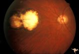
Bilateral Papilledema from Occipital Tumor | Left eye. Bilateral hemorrhagic papilledema. Occipital glioma. Woman. Anatomy: Optic disc. Pathology: Papilledema. Disease/Diagnosis: Hemorrhagic papilledema from occipital glioma. | Image |
| 163 |
 |
Bilateral Papilledema from Occipital Tumor | Right eye. Bilateral hemorrhagic papilledema. Occipital glioma. Right hemianopia. Woman. Anatomy: Optic disc. Pathology: Papilledema. Disease/Diagnosis: Hemorrhagic papilledema from occipital glioma. | Image |
| 164 |
 |
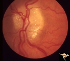
C105 Disc Edema with Systemic Lupus | Mild disc edema blurs the inferior disc margin. Flourescein angiogram in D1_06. Same patient as D1_06 an D1_07. Man. Anatomy: Optic disc. Pathology: Axoplasmic stasis due to vasculitis (Lupus). Disease/ Diagnosis: Lupus papillopathy. | Image |
| 165 |
 |
Chronic Atrophic Papilledema | Left eye. Left eye blind. Chronic Atrophic Papilledema. Obese woman (300 lbs) with large tentorial meningioma. "Pseudo Pseudotumor". Anatomy: Optic disc. Pathology: Papilledema. Disease/Diagnosis: Papilledema from large tentorial meningioma. | Image |
| 166 |
 |
Chronic Atrophic Papilledema | Right eye. Chronic Atrophic Papilledema. Obese woman (300 lbs) with large tentorial meningioma. "Pseudo Pseudotumor" Anatomy: Optic disc. Pathology: Papilledema. Disease/Diagnosis: Papilledema from large tentorial meningioma. | Image |
| 167 |
 |
Chronic Papilledema in Resolution. Sequence | Left eye 2 weeks after presentation. Chronic papilledema in resolution. Note first evidence of a vertical choroidal fold. Anatomy: Optic disc. Pathology: Papilledema. Disease/Diagnosis: Papilledema. | Image |
| 168 |
 |
Chronic Papilledema in Resolution. Sequence | Left eye at presentation. Chronic papilledema. Anatomy: Optic disc Pathology: Papilledema Disease/Diagnosis: Papilledema | Image |
| 169 |
 |
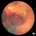
Chronic Papilledema with Hemorrhagic and Exudative Complications | Left eye one month after presentation. Resolving hemorrhage. Chronic papilledema with hemorrhagic and exudative complications due to Pseudotumor cerebri. Anatomy: Optic disc. Pathology: Papilledema. Disease/Diagnosis: Chronic papilledema with hemorrhagic and exudative complications | Image |
| 170 |
 |
Chronic Papilledema with Hemorrhagic and Exudative Complications | Left eye at presesntation. Chronic papilledema with hemorrhagic and exudative complications due to Pseudotumor cerebri. Anatomy: Optic disc Pathology: Papilledema Disease/Diagnosis: Chronic papilledema with hemorrhagic and exudative complications. | Image |
| 171 |
 |
Chronic Papilledema with Pseudo Drusen | Left eye. Chronic papilledema with pseudo drusen due to cerebral pontine angle tumor. Anatomy: Optic disc. Pathology: Papilledema Disease/Diagnosis: Chronic papilledema with pseudo drusen. | Image |
| 172 |
 |
Chronic Papilledema with Pseudo Drusen | Right eye. Chronic papilledema with pseudo drusen due to cerebral pontine angle tumor. Anatomy: Optic disc Pathology: Papilledema Disease/Diagnosis: Chronic papilledema with pseudo drusen | Image |
| 173 |
 |
Chronic Papilledema with Pseudo Drusen | Right eye. Meningioma. Pseudo drusen from chronic papilledema. Woman. Anatomy: Optic disc Pathology: Papilledema Disease/Diagnosis: Chronic papilledema with pseudo drusen | Image |
| 174 |
 |
Chronic Papilledema with Pseudo Drusen | Left eye of 51 year old, 220 pound black woman. Pseudotumor cerebri, pseudo drusen, exudates. Anatomy: Optic disc. Pathology: Papilledema Disease/Diagnosis: Chronic papilledema with pseudo drusen | Image |
| 175 |
 |
Chronic Papilledema with Pseudo Drusen | Chronic papilledema with pseudo drusen. Residual choroidal folds. Pseudo drusen. Anatomy: Optic disc. Pathology: Papilledema. Disease/Diagnosis: Chronic papilledema with pseudo drusen. | Image |