Best known for his world-renowned neuro-ophthalmology unit based at the University of California, San Francisco, William Hoyt, MD collected here more than 850 of his best images covering a wide range of disorders.
William F. Hoyt, MD, Professor Emeritus of Ophthalmology, Neurology and Neurosurgery, Department of Ophthalmology, University of California, San Francisco.
NOVEL: https://novel.utah.edu/
TO
Filters: Collection: ehsl_novel_wfh
| Identifier | Title | Description | Subject | ||
|---|---|---|---|---|---|
| 126 |
 |
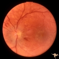
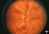
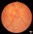
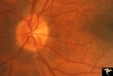
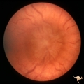
D1_04 | D104 Disc Edema with Systemic Lupus | Unilateral disc swelling and enlarged blind spot. Patient had episcleritis 4 weeks before this image was taken. 14 year old girl. Anatomy: Optic disc. Pathology: Axoplasmic stasis due to vasculitis (Lupus). Disease/ Diagnosis: Lupus papillitis. Clinical: No visual loss. History of episcleritis. Big ... | Disc Swelling; Inflammatory Papillopathies; Disc Edema; Systemic Lupus |
| 127 |
 |
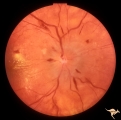
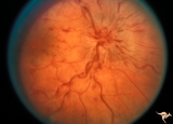
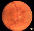
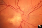
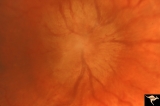
D1_05 | C105 Disc Edema with Systemic Lupus | Mild disc edema blurs the inferior disc margin. Flourescein angiogram in D1_06. Same patient as D1_06 an D1_07. Man. Anatomy: Optic disc. Pathology: Axoplasmic stasis due to vasculitis (Lupus). Disease/ Diagnosis: Lupus papillopathy. | Disc Swelling; Inflammatory Papillopathies; Disc Edema; Systemic Lupus |
| 128 |
 |
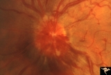
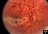
D1_06 | D106 Disc Edema with Systemic Lupus | Flourescein angiogram shows evidence of vascular papillopathy. (Lupus) Same patient as D1_05 an D1_07. Anatomy: Optic disc. Pathology: Axoplasmic stasis due to vasculitis (Lupus). Disease/ Diagnosis: Lupus papillopathy. | Disc Swelling; Inflammatory Papillopathies; Disc Edema; Systemic Lupus |
| 129 |
 |
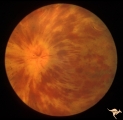
D1_07 | D107 Disc Edema with Systemic Lupus | Late stage Flourescein angiogram showing flourescein leakage on the disc and around the neighboring vessels. Note this amount of edema could not be appreciated in the colored fundus image D1_05. Same patient as D1_06 an D1_05. Anatomy: Optic disc. Pathology: Axoplasmic stasis due to vasculitis (Lupu... | Disc Swelling; Inflammatory Papillopathies; Disc Edema; Systemic Lupus |
| 130 |
 |
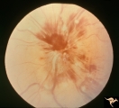
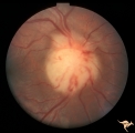
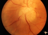
D2_01 | D201 Disc Edema with Systemic Hypertension | Left eye. Note generalized arterial narrowing. Low grade disc edema and multiple splinter hemorrhages. The patient had severe hypertension from kidney failure. Additional yellow intraretinal exudate at the macula. 20 year old male patient. Right eye. Pair with D2_02. Anatomy: Optic disc; Retina; Ret... | Disc Swelling; Inflammatory Papillopathies; Disc Edema; Systemic Hypertension |
| 131 |
 |
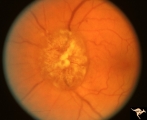
D2_02 | D202 Disc Edema with Systemic Hypertension | Right eye. Note generalized arteriole narrowing. Low grade disc edema and multiple splinter hemorrhages. The patient had severe hypertension from kidney failure. Additional yellow intraretinal exudate at the macula. 20 year old male patient. Pair with D2_01. Anatomy: Optic disc; Retina; Retinal art... | Disc Swelling; Inflammatory Papillopathies; Disc Edema; Systemic Hypertension |
| 132 |
 |
E01 | E01 Disc Swelling with Central Vein Occlusion | Left eye. Central retinal vein occlusion with disc swelling. Anatomyt: Optic disc; Retina. Pathology: Vasculitis. Disease/ Diagnosis: Disc swelling due to retinal vasculitis. | Disc Swelling; Central Vein Occlusion |
| 133 |
 |
E02 | E02 Disc Swelling with Central Vein Occlusion | 37 year old black male with sickle cell C causing unilateral central retinal vien occlusion. Anatomy: Optic disc; Retina. Pathology: Occlusion of the central retinal vein. Disease/ Diagnosis: Disc swelling due to central retial vein occlusion. Clinical: Visual blurring. | Disc Swelling; Central Vein Occlusion; Central Retinal Vein; Central and Branch Vein Occlusions |
| 134 |
 |
E03 | E03 Disc Swelling with Central Retinal Vein Occlusion | 36 year old woman with visual obscurations of right eye. Early CRVO, papillophlebitis. Steroid responsive. Anatomy: Optic disc; Retina. Pathology: Central retinal vein occlusion. Disease/ Diagnosis: Disc swelling due to central retinal vein occlusion. Clinical: Decreased vision in right eye. Acuity ... | Disc Swelling; Central Vein Occlusion |
| 135 |
 |
E04 | E04 Disc Swelling with Central Retinal Vein Occlusion | Acute CRVO, right eye with disc swelling. Male patient. Same patient as E05. Anatomy: Optic disc; Retina. Pathology: Central retinal vein occlusion. Disease/ Diagnosis: Disc swelling due to central retinal vein occlusion. Clinical: Visual blurring. | Disc Swelling; Central Vein Occlusion; Central Retinal Vein; Central and Branch Vein Occlusions |
| 136 |
 |
E05 | E05 Disc Swelling with Central Retinal Vein Occlusion | Resolving CVRO, right eye. Two months following slide E04. Male patient. Anatomy: Optic disc; Retina. Pathology: Central retinal vein occlusion. Disease/ Diagnosis: Resolved disc swelling after central retinal vein occlusion. Clinical: No symptoms. | Disc Swelling; Central Vein Occlusion |
| 137 |
 |
E06 | E06 Disc Swelling with Central Retinal Vein Occlusion | Acute disc swelling one week after onset of symptoms. Anatomy: Optic disc; Retina. Pathology: Central retinal vein occlusion. Disease/ Diagnosis: Disc swelling due to central retinal vein occlusion. Clinical: Visual blurring. | Disc Swelling; Central Vein Occlusion |
| 138 |
 |
E07 | E07 Disc Swelling with Central Vein Occlusion | 24 year old male. Papillophlebitis (CRVO) with optic disc edema. Right eye. Anatomy: Optic disc; Retina. Pathology: Central retinal vein occlusion. Disease/ Diagnosis: Disc swelling due to central retinal vein occlusion. Clinical: ??Branch retinal artery occlusion [sic]. | Disc Swelling; Central Vein Occlusion; Retinal Disorders; Vascular |
| 139 |
 |
E08 | E08 Disc Swelling with Central Vein Occlusion | Pituitary adenoma with right chronic CRVO with optociliary bypass vessels. Anatomy: Optic disc; Retina. Pathology: Central retinal vein occlusion. Disease/ Diagnosis: Disc swelling due to central retinal vein occlusion. | Disc Swelling; Central Vein Occlusion |
| 140 |
 |
E09 | E09 Disc Swelling with Central Vein Occlusion | Chronic disc swelling due to CRVO. Anatomy: Optic disc; Retina. Pathology: Central retinal vein occlusion. Disease/ Diagnosis: Disc swelling due to central retinal vein occlusion. | Disc Swelling; Central Vein Occlusion |
| 141 |
 |
E10 | E10 Disc Swelling with Central Vein Occlusion | Cilioretinal artery infarction after a central retinal vein occlusion. Anatomy: Optic disc; Retina. Pathology: Central retinal vein occlusion. Disease/ Diagnosis: Disc swelling due to central retinal vein occlusion. | Disc Swelling; Central Vein Occlusion; Retinal Disorders; Vascular |
| 142 |
 |
E11 | E11 Disc Swelling with Central Vein Occlusion | Old retinal vein occlusion with optociliary bypass vessel at 3:00. Right eye. Anatomy: Optic disc; Retina. Pathology: Central retinal vein occlusion. Disease/ Diagnosis: Disc swelling due to central retinal vein occlusion. | Disc Swelling; Central Vein Occlusion |
| 143 |
 |
E12 | E12 Disc Swelling with Central Vein Occlusion | 2nd attack of papillophlebitis. There is an optociliary bypass vessel at 4:00. Anatomy: Optic disc; Retina. Pathology: Central retinal vein occlusion. Disease/ Diagnosis: Disc swelling due to central retinal vein occlusion. | Disc Swelling; Central Vein Occlusion |
| 144 |
 |
F1_01 | F101 Optic Disc Lymphosarcoma | Optic disc lymphosarcoma. This disc has been infiltrated by neoplastic cells. Anatomy: Optic disc. Pathology: Lymphosarcoma. Disease/ Diagnosis: Lymphosarcoma. | Disc Swelling; Neoplastic Papillopathy; Metastatic Papillopathy |
| 145 |
 |
F1_02 | F102 Myeloblastic Leukemia | Myeloblastic leukemia. Left eye. Pair with F1_03. Anatomy: Optic disc. Pathology: Neoplastic (metastatic) papillopathy. Disease/ Diagnosis: Myeloblastic leukemia. | Disc Swelling; Neoplastic Papillopathy; Metastatic Papillopathy; Leukemias |
| 146 |
 |
F1_03 | F103 Myeloblastic Leukemia | Myeloblastic leukemia. Right eye. Pair with F1_02. Anatomy: Optic disc. Pathology: Neoplastic (metastatic) papillopathy. Disease/ Diagnosis: Myeloblastic leukemia. | Disc Swelling; Neoplastic Papillopathy; Metastatic Papillopathy; Leukemias |
| 147 |
 |
F1_04 | F104 Esthesio Neuroblastoma | Esthesioneuroblastoma. Tumor cells infiltrating the optic disc. 20/200 vision. Anatomy: Optic disc. Pathology: Esthesioneuroblastoma. Disease/ Diagnosis: Esthesioneuroblastoma. | Disc Swelling; Neoplastic Papillopathy; Metastatic Papillopathy |
| 148 |
 |
F1_05 | F105 Histiocytosis Infiltrate of Disc | Histiocytosis infiltrate of right disc with simultaneous infiltration of the hypothalamus with skin lesions on eye lids and chest. Same patient as F1_06. Anatomy: Optic disc. Pathology: Histiocytosis infiltrate. Disease/ Diagnosis: Histiocytosis infiltrate. Clinical: Patient presented with skin lesi... | Disc Swelling; Neoplastic Papillopathy; Metastatic Papillopathy; Histiocytoses |
| 149 |
 |
F1_06 | F106 Histiocytosis Infiltrate of Disc | More fully developed and chronic histiocytosis infiltrate of right disc with simultaneous infiltration of the hypothalamus with skin lesions on eye lids and chest. Same patient as F1_05, one year later. Anatomy: Optic disc. Pathology: Histiocytosis infiltrate of disc. Disease/ Diagnosis: Histiocytos... | Disc Swelling; Neoplastic Papillopathy; Metastatic Papillopathy; Histiocytoses |
| 150 |
 |
F1_07 | F107 Metastatic Breast Cancer to the Disc | Metastatic breast cancer to the disc. Notice mass on inferior portion of disc. Also notice tangled capillary dilation within the mass indicating infiltration. This disc tumor was radiated. It disappeared leaving a pale flat atrophic nerve. The patient died. Histologic study of the eye revealed metas... | Disc Swelling; Neoplastic Papillopathy; Metastatic Papillopathy; Metastatic Carcinoma |