The Health Education Assets Library (HEAL) is a collection of over 22,000 freely available digital materials for health sciences education. The collection is now housed at the University of Utah J. Willard Marriott Digital Library.
TO
Filters: Collection: ehsl_heal
| Title | Description | Subject | Collection | ||
|---|---|---|---|---|---|
| 126 |
 |
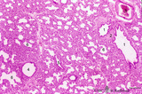
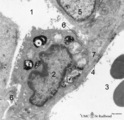
Survey of lung parenchym (gerbil) | Scanning electron microscopy. The photograph shows (1) bifurcation of several bronchi in the lung parenchym with alveoli (2). The long arrows indicate the route of alveolar ducts (4 ↕ length, and cross 4↑). Pulmonary vessels (3). | Lung parenchym; Pulmonary vessels | Poja Histology Collection - Respiratory System Subset |
| 127 |
 |
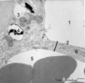
Survey of lung parenchym with bronchi and blood vessels (human, adult) | Stain: Azan. The bronchus (1) is surrounded with hyaline cartilage rings (2), seromucous glands (9) and muscle fibers (*), and lined with respiratory epithelium (←). Pulmonary arteries (3) in their neighborhood; a solitary pulmonary vein (8); (4) Carbon deposits of varying sizes (dust loaded macr... | Bronchiolus | Poja Histology Collection - Respiratory System Subset |
| 128 |
 |
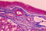
Survey of nose wing - human | Stain: Azan. At the top orofacial muscle (1, reddish) and hyaline cartilage (2, bluish) at the right. The external skin surface of the nose wing dot side (.) is covered with squamous epithelium, light-stained sebaceous glands (3) and hair structures (4). At the starred side (*) the nasal vestibulum... | Poja Histology Collection - Respiratory System Subset | |
| 129 |
 |
Survey of the nasal conchae (dog, isolated turbinate bones) | Stain: Hematoxylin and eosin. The conchae (turbinates) consist of three parts: the inferior, middle and superior turbinate bones (3, black-stained); they are covered by a respiratory mucosa (1) or by an olfactory mucosa (2). In humans only a small part of the superior concha exhibits olfactory epith... | Conchae nasales; Olfactory epithelium; Respiratory epithelium | Poja Histology Collection - Respiratory System Subset |
| 130 |
 |
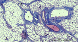
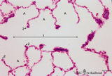
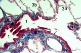
Terminal sac period of developing lung (human, fetus) | Stain: Hematoxylin and eosin. The transition of a terminal bronchiolus (1) into two future respiratory bronchioli (2), present as dilated spaces (saccules derived from the primitive respiratory channels, hence the name terminal sac period). The surrounding cellular tissue is composed of developing p... | Lung development; Terminal sac period; Respiratory bronchioli; Mesenchyme | Poja Histology Collection - Respiratory System Subset |
| 131 |
 |
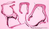
Terminal sac period of developing lung (human, fetus, low magnification) | Stain: Hematoxylin and eosin. At the right cross-sections of a large bronchus (1) with cartilage rings (2). Close to them a pulmonary artery (3), and a bronchiolus (4) without cartilage. (5) indicates a respiratory bronchiolus. Most alveoli are not inflated, the numerous dilated spaces (6) are saccu... | Lung development ; Terminal sac period; Respiratory bronchioli | Poja Histology Collection - Respiratory System Subset |
| 132 |
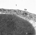 |
The air-blood barrier in the lung (rat) | Electron microscopy (high magnification). The alveolar space (1) and the capillary space (2) (with part of an erythrocyte, 3) are separated from each other respectively by cytoplasm of endothelial cell (5), the common basal lamina (6) with a lamina densa (6a) and the type I alveolar cell (4, pneumo... | Pneumocyte I; Alveolar cell type I | Poja Histology Collection - Respiratory System Subset |
| 133 |
 |
The alveolar tip in lung tissue (human, adult) | Electron microscopy. The lung tip is covered by a flattened alveolar cell type I (↓1) and a similar neighboring bulging one with nucleus (2). In the alveolar tip amorph elastin (5) appears more electron-dense than the collagen fibers (Col) and is interwoven with it. Small foci of calcifications a... | Alveolar tip; Elastoblast | Poja Histology Collection - Respiratory System Subset |
| 134 |
 |
The alveolus and air-blood barrier in the lung (rat) | Electron microscopy. The alveolus (1) is lined by a thin extension (2) of the alveolar epithelial cell type I (2), the pneumocyte I and the thin endothelium (3) of the capillary filled with erythrocytes (4) and blood platelets (5). The thin-walled air-blood barrier (↔) consists of the transition f... | Pneumocyte I; Alveolar cell type I | Poja Histology Collection - Respiratory System Subset |
| 135 |
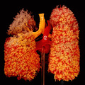 |
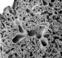
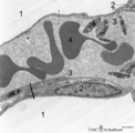
The bronchial tree and branches of the pulmonary artery (human, adult, posterior aspect) | Resin corrosion cast of left and right lung. Posterior aspect of lower trachea (1, yellow) with two principal bronchi (yellow) with red-stained pulmonary artery (2) and its branching. The cast of both lung lobes reveals especially the intricate divisions and branching of the bronchial tree (yellow) ... | Macroscopy | Poja Histology Collection - Respiratory System Subset |
| 136 |
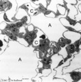 |
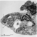
The terminal part of the airway: alveoli (dog) | Electron microscopy (low magnification). (A) indicate alveoli; (C) indicate capillaries. (1) type I alveolar cell (pneumocyte I, squamous alveolar cell); (2) type II alveolar cell (pneumocyte II, great alveolar cell). (3) alveolar macrophage. (4) endothelium of capillary. | Pneumocyte I; Pneumocyte II; Alveolar macrophage | Poja Histology Collection - Respiratory System Subset |
| 137 |
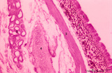 |
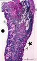
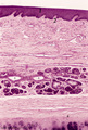
Trachea (cat) | Stain: Orcein and eosin. At the top pseudostratified epithelium (1) followed by a thick layer filled with dark-stained elastic fibers (2). Bundle of smooth muscle fibers (3) (note: there is an artifactual space between the elastic layer and the smooth muscle), belonging to the tunica fibro-musculo-... | Tracheal glands; Seromucous glands | Poja Histology Collection - Respiratory System Subset |
| 138 |
 |
Transition of respiratory bronchiolus into alveolar duct in the lung (human) | Stain: Azan. The alveolar duct (1↔) exhibits an alveolated wall but at a few places (3) bronchiolar characteristics are evident such as cuboidal epithelium covering small areas of smooth muscle and connective tissue. They differ from the characteristic alveolar tips (2↓) of neighboring thin-wall... | Alveolar ducts; Alveolar tips | Poja Histology Collection - Respiratory System Subset |
| 139 |
 |
Transition surface of vocal cord into laryngeal surface (human, subglottis, higher magnification) | Stain:Stain: Azan. Transition (↓) from stratified squamous epithelium (1) into pseudostratified epithelium (2) in the subglottis region. Blood vessels and the cellularity of the proper lamina is evident. | Olfactory epithelium; Subglottis; Stratified epithelium; Pseudostratified epithelium | Poja Histology Collection - Respiratory System Subset |
| 140 |
 |
Transition terminal bronchiolus - respiratory bronchiolus (human) | Stain: Azan. A longitudinal section shows the transition from the lumen of a terminal bronchiolus (1) into that of a respiratory bronchiolus (2). The epithelium of the terminal bronchiolus is regularly columnar and ciliated. The respiratory bronchiolus has an irregular lining and is covered by low c... | Terminal bronchiolus; Respiratory bronchiolus; Columnar epithelium; Cuboidal epithelium; Alveolar epithelium | Poja Histology Collection - Respiratory System Subset |
| 141 |
 |
Transitional region of epiglottis (human, high magnification) | Stain: Hematoxylin and eosin. Transition of squamous epithelium into pseudostratified epithelium. Note diapedesis of lymphocytes (darker stained rounded cells) through the epithelial barrier (→). The proper lamina presents some lymphocyte infiltration. | Diapedesis | Poja Histology Collection - Respiratory System Subset |
| 142 |
 |
Transitional zone in the middle part of nasal vestibulum (comparable with red zone or vermilion border of lip, human) | Stain: Azan. At the right slightly cornified squamous epithelium (1) with small dermal papillae (2) and numerous blood vessels (3). To the left the transition to pseudostratified epithelium (4). In the submucosa branching seromucous nasal glands with draining ducts (5). | Stratified epithelium; Pseudostratified epithelium; Nasal glands; Seromucous glands | Poja Histology Collection - Respiratory System Subset |
| 143 |
 |
Type I alveolar cell in the lung (human, adult) | Electron microscopy. At the top the alveolar space is lined by type I alveolar cell (1, pneumocyte I). The cytoplasm is well provided with organelles and few electron-dense lysosomes, and pinocytotic vesicles. These pneumocyte I cells cover the interalveolar septa that contain fibroblasts and bundle... | Pneumocyte I; Interstitium | Poja Histology Collection - Respiratory System Subset |
| 144 |
 |
Type II alveolar cell in the lung (rat) | Electron microscopy. At the top the alveolar space (1) is lined by a type II alveolar cell (pneumocyte II) (2) with blunt microvilli. In the cytoplasm the characteristic electron-dense multilamellar bodies (*) and light-stained swollen mitochondria are present. The lamellar bodies are responsible fo... | Pneumocyte I; Pneumocyte II; Alveolar cell type I; Alveolar cell type II; Multilamellar bodies; Interstitial cells | Poja Histology Collection - Respiratory System Subset |
| 145 |
 |
Type II and type I alveolar cells in the lung (human, adult) | Electron microscopy. At the top the alveolar space (1) is lined by a thin cytoplasm (2) of type I alveolar cell (pneumocyte I). At the left side part of a type II alveolar cell (3, pneumocyte II) with electron-dense remnants of two characteristic multilamellar bodies (4) as well as many organelles. ... | Pneumocyte I; Pneumocyte II; Interstitial cell; Alveolar cell type I; Alveolar cell type II | Poja Histology Collection - Respiratory System Subset |
| 146 |
 |
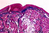
Upper part of nasal septum (dog, higher magnification) | Stain: Hematoxylin and eosin. Stratified squamous epithelium (1) with small papillae (2). The lamina propria contains many small and large thin-walled blood vessels also known as the venous plexus (sinusoids) of Auerbach (3, swell body). Nasal glands (4) with draining ducts (5, larger diameter) and ... | Squamous stratified epithelium; Nasal vestibulum; Venous sinusoids; Venous plexus; Swell bodies | Poja Histology Collection - Respiratory System Subset |
| 147 |
 |
Ventricular fold (plica ventricularis) (human) | Stain: Azan. On top pseudostratified epithelium (1). Lamina submucosa with predominantly mucous glands (3), fat cells (4) and blood vessels. The lamina propria also contains lymphoid follicles (2) with a germinal center (immune protection). | Laryngeal glands; Ventricular fold; Seromucous glands | Poja Histology Collection - Respiratory System Subset |
| 148 |
 |
Ventricular fold (plica ventricularis) (human, higher magnification) | Stain: Azan. On top pseudostratified columnar, ciliated epithelium (1). Centrally below an excretory duct (2) of laryngeal seromucous gland (3) with some adipocytes (*). Blood vessels and two lymph vessels (4) are present. | Supraglottis; Ventricular fold; Laryngeal glands; Seromucous glands | Poja Histology Collection - Respiratory System Subset |
| 149 |
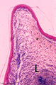 |
Vocal cord (plica vocalis) (human, glottis) | Stain: Azan. The true vocal cord consist of the vocal ligament (L) covered by stratified squamous epithelium and the striated vocal muscle (medial part of the thyreoarythenoid muscle, not depicted in this picture). At the tip of the vocal cord the stratified epithelium is distinct and the lamina pro... | Vocal ligament | Poja Histology Collection - Respiratory System Subset |