Best known for his world-renowned neuro-ophthalmology unit based at the University of California, San Francisco, William Hoyt, MD collected here more than 850 of his best images covering a wide range of disorders.
William F. Hoyt, MD, Professor Emeritus of Ophthalmology, Neurology and Neurosurgery, Department of Ophthalmology, University of California, San Francisco.
NOVEL: https://novel.utah.edu/
TO
Filters: Collection: ehsl_novel_wfh
| Title | Description | Type | ||
|---|---|---|---|---|
| 126 |
 |
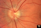
C106 Papillitis, Retrobulbar Neuritis | Papillitis with recovery of vision. Woman acupuncturist. Anatomy: Optic disc. Pathology: Axoplasmic stasis after inflammation. Disease/ Diagnosis: Optic neuritis/optic papillitis. Clinical: Visual loss with recovery. | Image |
| 127 |
 |
C107 Papillitis, Retrobulbar Neuritis | Man with bilateral papillitis. Right eye. Pair with C1_08. Cause unknown. Visual field showed central scotomas. Anatomy: Optic disc. Pathology: Axoplasmic stasis due to inflammation. Disease/ Diagnosis: Neuritis of the optic nerve. Clinical: Visual loss. | Image |
| 128 |
 |
C108 Papillitis, Retrobulbar Neuritis | Man with bilateral papillitis. Left eye. Pair with C1_07. Cause unknown. Visual field shows central scotoma. Anatomy: Optic disc. Pathology: Axoplasmic stasis due to inflammation. Disease/ Diagnosis: Optic neuritis / Optic papillitis. Clinical: Visual loss. | Image |
| 129 |
 |
C109 Papillitis, Retrobulbar Neuritis | Optic papillitis after wasp sting. 57 year old woman. Right eye. Anatomy: Optic disc. Pathology: Axoplasmic stasis due to inflammation. Disease/ Diagnosis: Optic neuritis after wasp sting. Clinical: Visual loss after wasp sting. | Image |
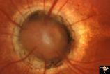
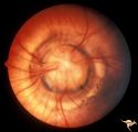
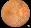
| 130 |
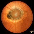 |
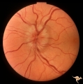
C11 Morning Glory Disc | "Morning Glory" disc. 11 year old girl. May not have a central retinal artery. Anatomy: Optic disc. | Image |
| 131 |
 |
C110 Papillitis, Retrobulbar Neuritis | AIDs papillitis. Segmental. Note inflammatory focus on temporal side of disc. 29 year old homosexual male. Visual field shows huge blind spot. Anatomy: Optic disc. Pathology: Axoplasmic stasis due to inflammation. Disease/ Diagnosis: AIDS papillitis / AIDS Optic neuritis. Clinical: Visual symptoms d... | Image |
| 132 |
 |
C111 Papillitis, Retrobulbar Neuritis | AIDS papillitis. Male. Anatomy: Optic disc. Pathology: Axoplasmic stasis due to inflammation. Disease/ Diagnosis: AIDS papillitis. Clinical: Visual symptoms. | Image |
| 133 |
 |
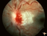
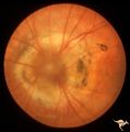
C112 Papillitis, Retrobulbar Neuritis | Woman with herpes. Acute retinal necrosis with papillitis and arcuate neuro-retinitis. Right eye. Pair with C1_13. Reference: Margolis T, Irvine AR, Hoyt WF, Hyman R. Acute retinal necrosis syndrome presenting with papillitis and arcuate neuroretinitis. Ophthalmology. 1988 Jul;95(7):937-40. Anatomy:... | Image |
| 134 |
 |
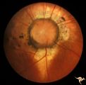
C113 Papillitis, Retrobulbar Neuritis | Woman with herpes. Acute retinal necrosis with papillitis an arcuate neuro-retinitis. Right eye. Notice the large arcuate defect extending fromt he disc to the retina of retinal necrosis. Pair with C1_12. Anatomy: Optic disc; Retina. Pathology: Axoplasmic stasis due to inflammation; Retinal necrosis... | Image |
| 135 |
 |
C114 Papillitis, Retrobulbar Neuritis | Epstien-Barr Virus papillitis with remarkably good recovery. Steroid responsive. Woman. Anatomy: Optic disc. Pathology: Axoplasmic stasis due to inflammation. Disease/ Diagnosis: Epstein Barr Virus with papillitis. Clinical: Visual loss that was steroid responsive. | Image |
| 136 |
 |
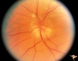
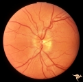
C115 Papillitis, Retrobulbar Neuritis | Demyelinative optic neuropathy with mild disc swelling. This eye had a large central scotoma. Note the bland disc margin swelling from 2:00 to 4:00. This swelling constitutes spill over edema from the main focus of the neuritis which lies behind the eyeball. Visual acuity was 2200. Anatomy: Optic di... | Image |
| 137 |
 |
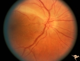
C116 Papillitis, Retrobulbar Neuritis | Demyelinative optic neuropathy with mild disc swelling. Note the mild disc margin blurring. The main focus of the neuritis lies behind the eye. 20/20 visual acuity. Anatomy: Optic disc; Optic nerve. Pathology: Axoplasmic stasis due to inflammation; Acute demyelination. Disease/ Diagnosis: Acute sub-... | Image |
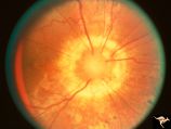
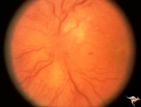
| 138 |
 |
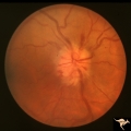
C12 Morning Glory Disc | "Morning Glory" disc. Patient 11 year old. Anatomy: Optic disc. | Image |
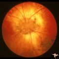
| 139 |
 |
C13 Morning Glory Disc | "Morning Glory" disc with peripapillary choroidal defect extending inferiorly. Patient has transphenoidal encephalocele. Note tapering edge like an arrow pointing to patient's basal encephalocele and cleft palate. Reference: Brodsky MC, Hoyt WF, Hoyt CS, Miller NR, Lam BL. Atypical retinochoroidal ... | Image |
| 140 |
 |
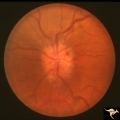
C14 Morning Glory Disc | Isolated "Morning Glory". Left eye. Girl. Anatomy: Optic disc. | Image |
| 141 |
 |
C15 Morning Glory Disc | "Morning Glory" disc. Note tapering edge pointing to patient's transphenoidal encephalocele. Reference: Brodsky MC, Hoyt WF, Hoyt CS, Miller NR, Lam BL. Atypical retinochoroidal coloboma in patients with dysplastic optic discs and transphenoidal encephalocele Arch Ophthalmol. 1995 May;113(5):624-8.... | Image |
| 142 |
 |
C16 Morning Glory Disc | "Morning Glory" disc. Note tapering edge pointing to basal encephalocele. Boy. Anatomy: Optic disc. | Image |
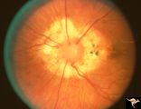
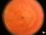
| 143 |
 |
C17 Morning Glory Disc | "Morning Glory" disc. CT normal. Anatomy: Optic disc. Clinical: CT normal. | Image |
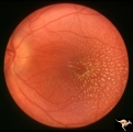
| 144 |
 |
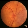
C18 Morning Glory Disc | "Morning Glory" disc. 6 month old baby. Anatomy: Optic disc | Image |
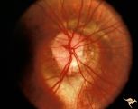
| 145 |
 |
C19 Morning Glory Disc | Bilateral "Morning Glory" disc. Right eye. Man. Pair with C_20. Anatomy: Optic disc. | Image |
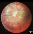
| 146 |
 |
C20 Morning Glory Disc | Bilateral "Morning Glory" disc. Left eye. Man. Pair with C_19. Anatomy: Optic disc. | Image |
| 147 |
 |
C201 Papillitis with Macular Star, Cat Scratch Disease | Proven Bartonella neuroretinitis. Notice the deposit of exudates of Henle's layer making an almost complete macular star. Anatomy: Optic disc; Retina. Pathology: Neuroretinitis; Axoplasmic stasis due to inflammation. Disease/ Diagnosis: Neuroretinitis due to Bartonella Henslae. Clinical: Visual blur... | Image |
| 148 |
 |
C202 Papillitis with Macular Star Cat Scratch Disease. | Proven Bartonella neuroretinitis. 23 year old man. Ocular disc edema with macular star (ODEMS). Anatomy: Optic disc; Retina. Pathology: Axoplasmic stasis due to inflammation; Exudate in Henle's layer. Neuroretinitis due to Bartonella Henslae (or cat scratch). Clinical: Visual blurring; Optic disc sw... | Image |
| 149 |
 |
C203 Papillitis with Macular Star, Cat Scratch Disease | Proven Bartonella neuroretinitis. Left eye. October 3, 1986. Same eye as C2_04. Macular star visible on C2_04. Woman. Ocular disc edema with macular star (ODEMS). Anatomy: Optic disc; Retina. Pathology: Axoplasmic stasis due to inflammation; Exudates in Henle's layer. Disease/ Diagnosis: Bartonella ... | Image |
| 150 |
 |
C204 Papillitis with Macular Star, Cat Scratch Disease | Proven Bartonella neuroretinitis. Left eye. October 17, 1986. Same eye as C2_03. Ocular disc edema with macular star (ODEMS). Woman. Anatomy: Optic disc; Retina. Pathology: Exudates in Henle's layer. DIsease/ Diagnosis: Neuroretinitis due to Bartonella Henslae (Cat Scratch). Clinical: Visual blurrin... | Image |