Best known for his world-renowned neuro-ophthalmology unit based at the University of California, San Francisco, William Hoyt, MD collected here more than 850 of his best images covering a wide range of disorders.
William F. Hoyt, MD, Professor Emeritus of Ophthalmology, Neurology and Neurosurgery, Department of Ophthalmology, University of California, San Francisco.
NOVEL: https://novel.utah.edu/
TO
| Title | Description | Type | ||
|---|---|---|---|---|
| 126 |
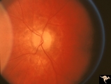 |
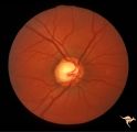
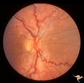
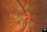
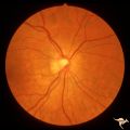
Resolution of Papilledema Following Optic Nerve Sheath Decompression (ONSD) | Left eye. 17 year old boy. Cryptococcal meningitis. Resolution of papilledema following optic nerve sheath decompression (ONSD) on November 1, 1974. Same eye as P_53a in January 1975. Atrophic, resolved disc. Note "high-water" marks. Visual acuity was 20/40. Anatomy: Optic disc. Pathology: Papilled... | Image |
| 127 |
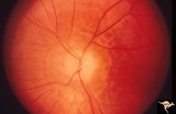 |
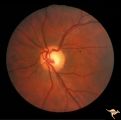
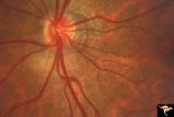
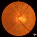
Resolution of Papilledema Following Optic Nerve Sheath Decompression (ONSD) | Left eye. 17 year old boy. Cryptococcal meningitis. Resolution of papilledema following optic nerve sheath decompression (ONSD) in November 1, 1974. Same eye as P_53a on December 1974. Atrophic. Note "high-water" marks. Anatomy: Optic disc. Pathology: Papilledema. Disease/Diagnosis: Resolving papill... | Image |
| 128 |
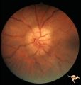 |
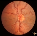
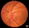
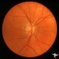
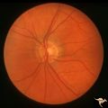
Bilateral Papilledema | Chronic Bilateral Papilledema. Anatomy: Optic disc. Pathology: Chronic bilateral papilledema. Disease/Diagnosis: Pseudotumor long standing. Clinical notes: Chronic headache; Obesity. | Image |
| 129 |
 |
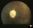
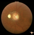
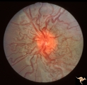
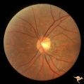
IC103a Central Retinal Artery Occlusion with Choroidal Arteriolar Occlusion | Central retinal artery occlusion and choroidal vascular occlusion due to pressure on the eyeball during craniotomy. Note total loss of vascularity of the optic disc and surrounding choroid. Anatomy: Optic disc. Pathology: Combined central retinal and choroidal arteriolar occlusion. Disease/ Diagnos... | Image |
| 130 |
 |
IC103d Central Retinal Artery Occlusion with Choroidal Arteriole Occlusion | 1969, Complete loss of blood supply to retina and choroid. Cause unknown. Boy. Anatomy: Optic disc. Pathology: Toxic ischemic retinal damage 22a. Clinical: Blindness. | Image |
| 131 |
 |
ID04b Post Papilledema Atrophy with Marked Gliosis | Post papilledema atrophy with marked gliosis in a patient with pseudotumor. Nasal ovoid absence of the retinal pigment epithelium. Presumably a defect from the long standing papilledema. 1985,. Right eye, pair with ID_4a. Anatomy: Optic disc. Pathology: Post papilledema atrophy and gliosis from long... | Image |
| 132 |
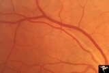 |
Ocular Hypertension | 1972. Right eye. Ocular hypertension. No field defect recognized. Pair with IIB1b & c. Anatomy: Peripapillary nerve fiber layer. Pathology: Slit-like defects in the arcuate nerve fiber bundles. Disease/Diagnosis: Elevated intraocular pressure. Clinical: Elevated intraocular pressure. | Image |
| 133 |
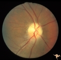 |
IB109 Post Ischemic (AION) Cupless Atrophy | Right eye, 1983 Top half of disc is pale. Striking focal arteriole narrowing. Anatomy: Optic disc. Pathology: Post ischemic (AION) cupless atrophy. Disease/ Diagnosis: Post ischemic (AION) cupless atrophy. Clinical: Visual loss. | Image |
| 134 |
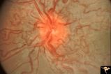 |
Retinocerebral Arteriovenous Malformation (Wyburn Mason Syndrome) | Florid arteriovenous malformation of the optic disc and surrounding retina, Caput medusa (Cross reference with V12-28 this section). Anatomy: Retina. Pathology: Arteriovenous malformation. Disease/Diagnosis: Wyburn Mason Syndrome. | Image |
| 135 |
 |
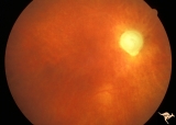
Sturge Weber Syndrome (Encephalotrigeminal Angiomatosis) | Sturge Weber Syndrome (Encephalotrigeminal angiomatosis); Color of the retina is deep red (sometimes called tomato catsup) due to a four fold thickening of the choroidal vascular bed. Optic disc is cupped due to elevated intraocular pressure. (Secondary glaucoma) Patient had a major ""port wine"" m... | Image |
| 136 |
 |
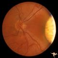
Sturge Weber Syndrome (Encephalotrigeminal Angiomatosis) | Left eye is normal, without the deep red from thickened Choroid. Pair with R1_B1a. Anatomy: Optic disc. Pathology: Diffuse choroidal hemangioma; Glaucoma. Disease/Diagnosis: Sturge Weber Syndrome. Clinical: Port wine hemangioma of the face. | Image |
| 137 |
 |
Vascular Disc Anomalies - Retinal Arteriovenous Malformations | Retinal arteriovenous malformations. No corresponding malformation of brain. Anatomy: Optic disc. Pathology: Retinal arteriovenous malformation. Disease/Diagnosis: Retinal arteriovenous malformation. Clinical: Asymptomatic. | Image |
| 138 |
 |
Venous Anomalies - Congenital Venous Tortuosity | Congenital venous tortuosity. Left eye. 9 year old boy. Same patient as V_51. Anatomy: Optic disc. Pathology: Congenital venous tortuosity. Disease/Diagnosis: Congenital venous tortuosity. Clinical: Asymptomatic. | Image |
| 139 |
 |
Venous Anomalies - Congenital Venous Tortuosity | Congenital venous tortuosity. Right eye. 9 year old boy. Same patient as V_52. Anatomy: Optic disc. Pathology: Congenital venous tortuosity. Disease/Diagnosis: Congenital venous tortuosity. Clinical: Asymptomatic. | Image |
| 140 |
 |
IC103c Central Retinal Artery Occlusion with Choroidal Arteriole Occlusion | 1988, Central retinal artery occlusion and choroidal vascular occlusion, 70 year old woman with history of central retinal artery occlusion 30 years prior. Anatomy: Optic disc. Pathology: Combined central retinal and choroidal arteriolar occlusion. Disease/ Diagnosis: Combined central retinal and ch... | Image |
| 141 |
 |
B106 Disc Swelling, Ischemic Papillopathies, AION | Red ischemic swelling. 49 year old man. Anatomy: Optic disc. Pathology: Axoplasmic stasis due to ischemia. Disease/ Diagnosis: AION. Clinical: Visual loss. | Image |
| 142 |
 |
H35 Segmental Hypoplasia, Retinal-Congenital Toxo | Left eye. Moving out temporally to see large chorioretinal scar. Temporal sector hypoplasia from congenital retinal toxoplasmosis. Same patient as H_34. Anatomy: Optic disc; Retina. Pathology: Hypoplasia secondary to retinal lesion. Disease/ Diagnosis: Segmental optic disc hypoplasia | Image |
| 143 |
 |
H48 Segmental Hypoplasia, Retinal-Nasal Hypoplasia | Bilateral nasal hypoplasia with absence of nasal nerve fiber layer and corresponding flag-like temporal field defect. Right eye. Same patient as H_49. Anatomy: Optic disc. Pathology: Nasal segmental disc hypoplasia. Disease/ Diagnosis: Congential anomaly. | Image |
| 144 |
 |
H49 Segmental Hypoplasia, Retinal-Nasal Hypoplasia | Bilateral nasal hypoplasia with absence of nasal nerve fiber layer and corresponding flag-like temporal field defect. Left eye. Same patient as H_48. Anatomy: Optic disc. Pathology: Nasal segmental disc hypoplasia. Disease/ Diagnosis: Congential anomaly. | Image |
| 145 |
 |
H46 Segmental Hypoplasia, Retinal-Nasal Hypoplasia | Nasal hypoplasia with suspected nasal pit about 3:00. Right eye. Man with temporal sector field defect. Same patient as H_47. Anatomy: Optic disc. Pathology: Nasal segmental disc hypoplasia. Disease/ Diagnosis: Congenital anomaly | Image |
| 146 |
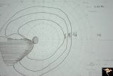 |
H42 Segmental Hypoplasia, Retinal, Nasal Hypoplasia | Visual field of patient in H_43. Flag-like temporal field defect from patient with nasal segmental disc hypoplasia. Anatomy: Optic disc. Pathology: Nasal segmental disc hypoplasia. Disease/ Diagnosis: Congenital anomaly. | Image |
| 147 |
 |
H44 Segmental Hypoplasia, Retinal, Nasal Hypoplasia | Bilateral nasal hypoplasia with bilateral flag-like temporal field defect. Right eye. Same patient as H_45. Anatomy: Optic disc. Pathology: Nasal segmental disc hypoplasia. Disease/ Diagnosis: Congenital anomaly. | Image |
| 148 |
 |
H45 Segmental Hypoplasia, Retinal-Nasal Hypoplasia | Bilateral nasal hypoplasia with bilateral flag-like temporal field defect. Left eye. Same patient as H_44. Anatomy: Optic disc. Pathology: Nasal segmental disc hypoplasia. Disease/ Diagnosis: Congenital anomaly. | Image |
| 149 |
 |
H47 Segmental Hypoplasia, Retinal-Nasal Hypoplasia | Normal left eye. Same patient as H_46. Anatomy: Optic disc. Pathology: Nasal segmental disc hypoplasia. Disease/ Diagnosis: Congenital anomaly. | Image |
| 150 |
 |
H43 Segmental Hypoplasia, Retinal, Nasal Hypoplasia | Nasal hypoplasia barely perceptible on disc. Nasal retinal nerve fibers are completely absent from 7:00 - 12:00. Anaotmy: Optic disc. Pathology: Nasal segmental disc hypoplasia. Disease/ Diagnosis: Congenital anomaly. | Image |