Collection of materials relating to neuro-ophthalmology as part of the Neuro-Ophthalmology Virtual Education Library.
NOVEL: https://novel.utah.edu/
TO
- NOVEL230
| Title | Creator | Description | Subject | ||
|---|---|---|---|---|---|
| 126 |
 |
Introduction to Examination of the Vestibular System | Daniel R. Gold, DO | Introduction to the Vestibular System Examination section of the NExT curriculum. | Exams; Vestibular System |
| 127 |
 |
Optical Coherence Tomography Angiography | David Zhao; Amanda Henderson, MD | Video presentation covering a thorough overview of Optical Coherence Tomography Angiography (OCTA). | Optical Coherence Tomography Angiography; OCTA |
| 128 |
 |
Introduction to Diagnostic Testing: Audio-Vestibular | Daniel R. Gold, DO | Introduction to the audio-vestibular diagnostic testing section of the NExT curriculum. | Diagnostic Testing; Exams; Audio-Vestibular |
| 129 |
 |
Ocular Lateropulsion Left AICA Stroke | Jorge C Kattah, MD | 82 year-old patient with basilar artery stenosis, she developed an acute left AICA stroke. On examination within 24 hours from symptom onset, she had primary gaze, unidirectional, right beat nystagmus and a positive left head impulse test. Brief periods of eyelid closure were associated with a h-... | Ocular Lateropulsion; AICA Stroke |
| 130 |
 |
Postconcussion Syndrome and Postconcussion Headache | Jessica Darusz, MD; Sean Gratton, MD | This brief presentation describes the pathophysiology, evaluation, and management of concussion, with an emphasis on postconcussion headache. | Concussion; Postconcussion Syndrome; Postconcussion Headache |
| 131 |
 |
Temporal Artery Biopsy Procedure | Nooran Badeeb; Danah Albreiki | Temporal artery biopsy is a procedure that is done in a patient with suspicion of GCA (Giant cell Arteritis), and some of the clinical manifestations that prompts us to suspect the diagnosis in patients older than 50 years old are: 1. GCA symptoms e.g. new onset headache. 2 . Visual symptoms: - Visi... | Temporal Artery Biopsy; GCA; Temporal Arteritis |
| 132 |
 |
Intraocular Pressure (IOP) Measurement: A Simple Guide | Nandini Singh; Kirsty Sumerville Mcalester; Alicia Yap; Anne Lee | This video demonstrates the technique for measuring intraocular pressure (IOP) and the use of the tonopen. | Intraocular Pressure (IOP); Tonometry |
| 133 |
 |
Selective Saccadic Palsy After Cardiac Surgery | Nilan D. Schnure, MD; Ali G. Hamedani, MD, MHS; Grant T. Liu, MD | A unique gaze palsy selectively affecting saccades while sparing smooth pursuit, vergence, and vestibular reflex eye movements has been described following uncomplicated cardiac (and especially ascending aortic) surgery . This 69 year-old man reported persistent visual complaints immediately after a... | Saccade; Saccadic Palsy; Cardiac Surgery |
| 134 |
 |
An Introduction to CT and MRI in Neuro-Imaging | Michael Carper, MD | A brief lecture covering basic neuro-imaging, including computed tomography (CT) and magnetic resonance imaging (MRI). | Computed Tomography (CT); Magnetic Resonance Imaging (MRI); Neuro-imaging |
| 135 |
 |
Visual Field Testing in the Pediatric Population | Lauren C. Ditta, MD | Visual field testing can be performed in the pediatric population with ease, however the proper technique for testing must be utilized and is tailored to each child's unique clinical situation and age. Confrontation visual field testing is a quick and easy; test that can yield a rough assessment of ... | Visual Field Testing; Pediatric; Confrontation Visual Field; Automated; Perimetry |
| 136 |
 |
Introduction to the Pupil Examination | Karl C. Golnik, MD | Introduction to pupil examination techniques. | Pupil Examination |
| 137 |
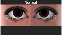 |
Normal Light Reflex and Relative Afferent Pupillary Defect (RAPD) | Marshall Huang, 4th Year Medical Student | A Relative Afferent Pupillary Defect is an examination finding in patients who have an asymmetric pupillary reaction to light when it is shined back and forth between the two eyes. It is most commonly a sign of asymmetric optic nerve disease or damage but can also present in widespread asymmetric r... | Light Reflex; RAPD |
| 138 |
 |
Pendular vs Jerk Nystagmus | Tony Brune, DO; Daniel R. Gold, DO | A video distinguishing pendular and jerk nystagmus. | Nystagmus; Pendular Nystagmus; Jerk Nystagmus |
| 139 |
 |
Sensory Nystagmus | Tony Brune, DO; Jonathan D. Trobe, MD; Raed Behbehani, MD | A video describing sensory nystagmus. | Nystagmus; Sensory Nystagmus |
| 140 |
 |
Introduction to Examination of the Orbit and the Extraocular Structures in NANOS NOTE | Julie Falardeau, MD | Introduction to Examination of the Orbit and the Extraocular Structures | Orbit; Anatomy |
| 141 |
 |
Headache Associated With Low or High Intracranial Pressure | Yu Hsin Chen; Amanda Dean Henderson; James Brian Davis | When intracranial pressure changes, the most common symptom is headache, which often has a positional component. We introduce various etiologies of headaches attributed to increased or low intracranial pressure, with idiopathic intracranial hypertension (or primary pseudotumor cerebri), post-dural p... | CSF Leak; Headache; Intracranial Hypertension; Intracranial Hypotension; Papilledema; Pseudotumor Cerebri |
| 142 |
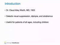 |
Worth 4-Dot Test | Angela Cao; Anne Abel | This presentation describes the Worth 4-Dot test, demonstrates how to perform the test, and interpret results. | Amblyopia; Diplopia; Worth 4-dot Test |
| 143 |
 |
Vogt-Koyanagi-Harada Syndrome | Shwetha Mudalegundi, Medical Student; Amanda D. Henderson, MD | Vogt-Koyanagi-Harada (VKH) syndrome is a rare disorder that affects several body systems. Here we take a broad look at the presentation and pathophysiology of VKH, with a more specific focus on the relevant eye findings. Since much is not known about VKH, we explore the current standards for diagnos... | Vogt-Koyanagi-Harada Syndrome; Uveitis |
| 144 |
 |
Sports Related Head Injuries | Jessica Darusz, MD; Sean Gratton, MD | This narrated PowerPoint reviews the basics of sports related head injuries. It emphasizes assessment tools and treatment decisions in assessing athletes with concussion and other head injuries. | Concussion; Sports-related Head Injuries; Post-concussion Syndrome |
| 145 |
 |
NANOS Illustrated Curriculum Section H Introduction | Sean Gratton, MD; Zoë R. Williams, MD | Introduction video to the resources available in Section H of the NANOS Illustrated Curriculum. | History, Neuro-ophthalmology; Principles of Practice, Neuro-Ophthalmology; Systems of Healthcare; Research, Neuro-ophthalmology |
| 146 |
 |
NANOS Illustrated Curriculum Section D Introduction | Meagan D. Seay, DO; Victoria S. Pelak, MD | Introduction video to the resources available in Section D of the NANOS Illustrated Curriculum. | Lesions, Afferent Visual Pathway; Disorders, Afferent Visual Pathway; Pupillary Anatomy; Disorders, Pupillary Function; Lesions, Efferent Visual Pathway; Ocular Motility; Abnormal Eye Movements; Eyelids; Abnormal Facial Movements |
| 147 |
 |
NANOS Illustrated Curriculum Section G Introduction | Meagan D. Seay, DO | Introduction video to the resources available in Section G of the NANOS Illustrated Curriculum. | Obesity, Management; Low Vision, Management; Genetic Diseases, Management; Patient Counseling |
| 148 |
 |
Introduction to Ocular Examination | Ore-ofe Adesina, MD | Introduction to the Ocular Examination section of the NExT curriculum. | Exams; Ocular Examination |
| 149 |
 |
Introduction to the Neurological Examination in NANOS NOTE | Padmaja Sudhakar, MD | An introduction to the neurological examination. | Neurology; Neurological Examination |
| 150 |
 |
Diagnosis and Evaluation of Stroke: Cerebral Hypoxia | Danny Alevy; James Brian Davis; Amanda Dean Henderson | Neurons are particularly vulnerable to oxygen deprivation. Cerebral hypoxia can be caused by arterial thrombosis, embolism, hypoperfusion, cervical artery dissection, or cryptogenic causes. About 1/3 of ischemic strokes are from cryptogenic causes. Embolic strokes are the next most common (20-30%), ... | Atherosclerosis; Cerebral Hypoxia; Cervical Artery Dissection; Cryptogenic Ischemia; Embolism; Hypoperfusion; Ischemia; Stroke; Thrombosis |