John A. Moran Eye Center Neuro-Ophthalmology Collection: A variety of lectures, videos and images relating to topics in Neuro-Ophthalmology created by faculty at the Moran Eye Center, University of Utah, in Salt Lake City.
NOVEL: https://novel.utah.edu/
TO
Filters: Collection: "ehsl_novel_jmec"
| Title | Description | Type | ||
|---|---|---|---|---|
| 126 |
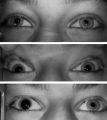 |
Left-sided Horner's Syndrome with an Acquired Preganglionic Localization | Left-sided Horner's syndrome in a 12-year-old girl with an acquired preganglionic localization based on clinical and pharmacologic testing. The cause remained undetermined after extensive radiologic investigations. Left-sided ptosis and miosis are evident in room light (top), and the degree of aniso... | Image |
| 127 |
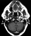 |
Left-sided Internal Carotid Artery Dissection | Left-sided internal carotid artery dissection identified on T-1 weighted magnetic resonance image from a 52-year-old man who suddenly developed left-sided neck and orbital pain along with a droopy left upper eyelid while dragging a deer out of the woods during hunting season. The normal dark flow vo... | Image |
| 128 |
 |
Levator Disinsertion | Example of patient with levator disinsertion, a lid disorder. Patient is pregnant and wears poorly fitting contacts. Discussion of characteristics, such as lid ptosis (shown in the left eye of patient), but with full levator function. | Image/MovingImage |
| 129 |
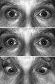 |
Light-near Dissociation | Light-near dissociation in a 51-year-old woman with multiple sclerosis who experienced double vision for 1 week. Her pupils are 5 mm in diameter in room light (top), react poorly in response to direct light reaction (middle), but constrict promptly in response to near stimulation (bottom). She also ... | Image |
| 130 |
 |
Light-near Dissociation | Example of patient with Argyll Robertson pupil with neurosyphilis. Shows a lack of pupillary response to light and some pupillary response to nearness of finger. | Image/MovingImage |
| 131 |
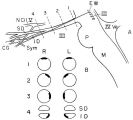 |
Location of Pupillomotor Fibers | Location of pupillomotor fibers are depicted as dark regions on cross-sections of the right (R) and left (L) oculomotor nerve at various locations along its course, including its emergence from the brain stem in the interpeduncular fossa (1), the midsubarachnoid segment (2), the level of the dorsum ... | Image |
| 132 |
 |
MELAS and RP | MELAS; Mitochondrial Encephalopathy with Lactic Acidosis, Stroke and Pigmentary Changes in retina-associated with a retinal dystrophy. This 53 year old man had seizures, encephalopathy and lactic acidosis typical of MELAS. His fundus examination showed granularity and some slight pigmentary changes ... | Text |
| 133 |
 |
Macula | Overview of the structure and viewing of the macula. | Text |
| 134 |
 |
Marcus Gunn Jaw Winking | Example of patient with Marcus Jaw Winking. Patient is led through instructions for movement of jaw (open, close, back and forth), with eyelid seen to be affected. Patient is then led through instructions for direction of gaze and pursuit. | Image/MovingImage |
| 135 |
 |
Measuring Visual Acuity | Demonstration on self of visual acuity exam, using a standard card. | Image/MovingImage |
| 136 |
 |
Migraine and Cluster Pathophysiology and Treatment | Video lecture covering pathophysiology and treatment of migraine and cluster headaches by Kathleen Digre, MD. | |
| 137 |
 |
Mimics of Atrophy | Text | |
| 138 |
 |
Monocular Pendular Nystagmus | Example of a patient with monocular pendular nystagmus, with discussion of situations in which this condition is seen: acquired disorder of the visual-sensory pathway, and acquired disorder of the brain stem (e.g. multiple sclerosis). | Image/MovingImage |
| 139 |
 |
Multifocal Electroretinograms | The most important development in ERGs is the multifocal ERG (mfERG). Erich Sutter adapted the mathematical sequences called binary m-sequences creating a program that can extract hundreds of focal ERGs from a single electrical signal. This system allows assessment of ERG activity in small areas of ... | |
| 140 |
 |
Near Reflex and Accomodation | Description of testing the near reflex and accomodation. | |
| 141 |
 |
Normal Eye Movements | This is an examination of a person with normal eye movements. Notice the patient has normal excursions. He has normal pursuit and saccades (horizontally and vertically). | Text |
| 142 |
 |
Normal Light Reflex without RAPD | This clip demonstrates the examination of the Relative Afferent Pupillary Defect (RAPD.) Demonstration of gauging the size of the pupil in light, testing light reflexes, swinging flashlight test for optic nerve abnormality. | Image/MovingImage |
| 143 |
 |
Normal Optic Disc | Overview of the structure and function of the normal optic disc. | Text |
| 144 |
 |
Notching of the Neuro-retinal Rim | The neuro-retinal rim becomes thinner; in particular the rim superotemporally and inferortemporally may develop a notch which is usually superior or inferior and rarely nasal or temporal. These notches are believed to be due to focal ischemic damage to the neuro-retinal rim. Glaucoma with Notching a... | Image |
| 145 |
 |
Nutritional Amblyopia | Example of patient with amblyopia with nutritional causes. | Text |
| 146 |
 |
Ocular Flutter | Two examples of patients, the first with rotary, flutter-like movements, but not ocular flutter, and the second with genuine ocular flutter. Discussion of difference between ocular flutter and nystagmus, and how to elicit ocular flutter. | Image/MovingImage |
| 147 |
|
Ocular Lateropulsion (Wallenberg's Syndrome) | Example of patient with ocular lateropulsion. Patient also has central Horner syndrome and nystagmus in right gaze. When shifting gaze back to forward, eyes overshoot their mark. Eyes laterally deviate to the right upon opening. | Image/MovingImage |
| 148 |
 |
Ocular Myasthenia | Example of patient with myasthenia gravis. Demonstration of tensilon test. Patient shown to have bilateral ptosis, bilateral duction deficits, and left hypertropia. Discussion of techniques to observe subtle changes, such as bringing in a neutral observer or taking still photographs. Shows split-scr... | Image/MovingImage |
| 149 |
 |
Ocular Myotonia | Example of patient with ocular myotonia. Patient is led through instructions for direction of gaze and opening and closing of eyes. Right eye is shown to be stuck in position after held gaze to the left and right, with very slow relaxation back into forward gaze. | Image/MovingImage |
| 150 |
 |
Oculopalatal Myoclonus | Oculopalatal myoclonus (OPM) Rhythmic oscillations of eyes and palate. Occurred after specific brainstem injury from stroke, following stenting. Related PowerPoint Presentation: http://content.lib.utah.edu/u?/EHSL-Moran-Neuro-opth,129 Disease/Diagnosis: Oculopalatal myoclonus. | Image/MovingImage |