John A. Moran Eye Center Neuro-Ophthalmology Collection: A variety of lectures, videos and images relating to topics in Neuro-Ophthalmology created by faculty at the Moran Eye Center, University of Utah, in Salt Lake City.
NOVEL: https://novel.utah.edu/
TO
Filters: Collection: "ehsl_novel_jmec"
| Title | Description | Type | ||
|---|---|---|---|---|
| 126 |
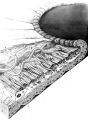 |
Structures of the iris | Structures of the iris. The a indicates the anterior border layer that terminates at the pigmentary ruff of the pupillary border (b). The c indicates the iris sphincter muscle, which is oriented circumferentially within the stroma and located deep to the anterior border layer; d indicates vessels th... | Image |
| 127 |
 |
Dysthyroid Optic Neuropathy: A Preventable Cause of Blindness | Dysthyroid Optic Neuropathy (DON) is a treatable cause of visual loss in ~5% of pts w/ ted. Monitor closely those pts with risk factors (proptosis, tight orbit, restricted motility, strabismus, smoker, diabetic). Oral prednisone is often effective, but frequent relapses after tapering. Orbital xrt ... | |
| 128 |
 |
See-saw Nystagmus | 7-year-old female whose mother noticed her eyes "bouncing" for 2 months. Visual acuity 20/70 OD and 20/40 OS, reduced color vision OU, and no afferent pupillary defect. See-saw nystagmus documented with videography. Manual perimetry revealed a complete right homonymous hemianopia. MRI revealed a lar... | Image/MovingImage |
| 129 |
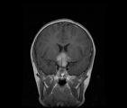 |
See-saw Nystagmus MRI 1 | MRI; See-saw Nystagmus | Image |
| 130 |
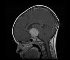 |
See-saw Nystagmus MRI 2 | MRI; See-saw Nystagmus | Image |
| 131 |
 |
Bilateral Asynchronous Blepharospasm with Facial and Cervical Dystonia | Bilateral Asynchronous Blepharospasm with Facial and Cervical Dystonia. | Image/MovingImage |
| 132 |
 |
Oculopalatal Myoclonus | Oculopalatal myoclonus (OPM) Rhythmic oscillations of eyes and palate. Occurred after specific brainstem injury from stroke, following stenting. Related PowerPoint Presentation: http://content.lib.utah.edu/u?/EHSL-Moran-Neuro-opth,129 Disease/Diagnosis: Oculopalatal myoclonus. | Image/MovingImage |
| 133 |
 |
Oculopalatal Myoclonus (PPT) | Oculopalatal myoclonus (OPM) Rhythmic oscillations of eyes and palate. Occurred after specific brainstem injury from stroke, following stenting. Related Video: http://content.lib.utah.edu/u?/EHSL-Moran-Neuro-opth,128 Disease/Diagnosis: Oculopalatal myoclonus | Image/MovingImage |
| 134 |
 |
Duane's Syndrome Type 1 | Clip of patient with Duane's Syndrome Type I. Presented at the Neurology Grand Rounds in Fall 2011 at the University of Utah. Presentation can be found in this collection at: Why Don't You See Double? http://content.lib.utah.edu/u?/EHSL-Moran-Neuro-opth,132 Disease/Diagnosis: Duane's Syndrome Type ... | Image/MovingImage |
| 135 |
 |
Duane's Syndrome Type 3 | Clip of patient with Duane's Syndrome Type III. Presented at the Neurology Grand Rounds in Fall 2011 at the University of Utah. Presentation can be found in this collection at: Why Don't You See Double? http://content.lib.utah.edu/u?/EHSL-Moran-Neuro-opth,132 Disease/Diagnosis: Duane's Syndrome Ty... | Image/MovingImage |
| 136 |
 |
Why Don't You See Double? | This presentation was given at the Neurology Grand Rounds in Fall 2011 at the University of Utah. A number of Duane Syndrome cases are covered. Related video can be found in this collection at: Duane's Syndrome Type I: http://content.lib.utah.edu/u?/EHSL-Moran-Neuro-opth,130 Duane's Syndrome Type I... | Text |
| 137 |
 |
B-scan Technique | This video describes and demonstrates the B-scan examination technique for examination of the eye using ultrasonography. | Image/MovingImage |
| 138 |
 |
Ultrasonography Techniques | This video describes and demonstrates the various techniques for examination of the eye using ultrasonography, including A-scan, B-scan and immersion. | Image/MovingImage |
| 139 |
 |
Ultrasonography: Immersion Technique | This video describes and demonstrates the immersion technique for examination of the eye using ultrasonography. | Image/MovingImage |
| 140 |
 |
The Orbital Exam | Comprehensive demonstration of the entire orbital examination. | |
| 141 |
 |
Fluoresein Angiography | Comprehensive description of using fluoresein angiography in examinations. | |
| 142 |
 |
Silent Sinus Syndrome | Silent sinus syndrome (SSS) is characterized by spontaneous and progressive unilateral enophthalmos. | |
| 143 |
 |
Benign Episodic Unilateral Mydriasis | Presentation covering benign episodic mydriasis. | Text |
| 144 |
 |
Clover-leaf Visual Field Defects | Description of clover-leaf visual field defects. | |
| 145 |
 |
The Wall-Eyed Potato Farmer | Young man presenting with apparent episodic neurologic evants that initially was thought to be multiple sclerosis, but as time went on, he had progressive changes in his neurologic exam and in his imaging findings. Brain biopsy revealed Gliomatosis cerebri. Anatomy: Brain Stem; Pons; Midbrain. Patho... | |
| 146 |
 |
Introduction to Fogging Refraction | An introduction to fogging refraction. | |
| 147 |
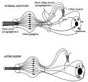 |
Pathophysiology of Signs Associated with a Tonic Pupil | Pathophysiology of signs associated with a tonic pupil. Normally, all parasympathetic fibers of the third cranial nerve synapse in the ciliary ganglion (top). Most postganglionic fibers innervate the ciliary muscle (dashed lines). After injury to the ciliary ganglion, the pupil becomes denervated an... | Image |
| 148 |
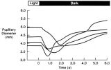 |
Pupillogram Demonstrating Paradoxical Pupillary Constriction to Darkness | Pupillogram demonstrating paradoxical pupillary constriction to darkness in four patients with congenital achromatopsia. Note that the pupils initially constrict when the light is extinguished. (Price MJ, Thompson HS, Judisch GF et al: Pupillary constriction to darkness. Br J Ophthalmol 1981;69:205-... | Image |
| 149 |
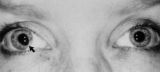 |
Tadpole-shaped Pupil | Tadpole-shaped pupil in a 20-year-old women with frequent episodes of blurred vision and achiness of the right eye lasting several minutes. The patient took a photograph of her eyes during an attack to document the peaked, segmental dilation of her right pupil (black arow). (Thompson HS, Zackon DH, ... | Image |
| 150 |
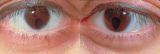 |
Bilateral Iris Colobomas | Coloboma literally means a "gap"-and can be used to describe any fissure, hole, or gap in the eye. The term most often is used to refer to a congenital gap in the disc, retina, the choroid, and the iris. Colobomas occur because the embryonic fissure fails to fuse. Since the fissure closure begins in... | Image |