A collection of videos relating to the diagnosis and treatment of eye movement disorders. This collection includes many demonstrations of examination techniques.
Dan Gold, D.O., Associate Professor of Neurology, Ophthalmology, Neurosurgery, Otolaryngology - Head & Neck Surgery, Emergency Medicine, and Medicine, The Johns Hopkins School of Medicine.
A collection of videos relating to the diagnosis and treatment of eye movement disorders.
NOVEL: https://novel.utah.edu/
TO
Filters: Collection: "ehsl_novel_gold"
| Title | Description | Type | ||
|---|---|---|---|---|
| 126 |
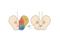 |
Figure 65: Vascular Distribution and Anatomy (Including 3rd Nerve) of the Rostral Midbrain | In this axial section of the midbrain at the level of the superior colliculus, the paired 3rd nuclei are located ventral to the periaqueductal grey, and the midline central caudal nucleus (CCN) is located in between. The fascicles that exit the IIIrd nuclei carry the fibers destined to innervate the... | Image |
| 127 |
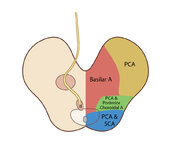 |
Figure 65: Vascular Distribution and Anatomy (Including 3rd Nerve) of the Rostral Midbrain (Supplement) | Image | |
| 128 |
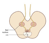 |
Figure 65: Vascular Distribution and Anatomy (Including 3rd Nerve) of the Rostral Midbrain (Supplement) | Image | |
| 129 |
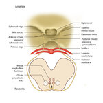 |
Figure 68: The Course of the 4th (IV) Nerve | The 4th nucleus lies at the ventral border of the periaqueductal gray matter, at the level of the inferior colliculus. The fascicles exit the nucleus dorsally and decussate at the anterior medullary velum (anterior floor of the fourth ventricle), and then exit the brainstem dorsally. The peripheral ... | Image |
| 130 |
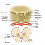 |
Figure 68: The Course of the 4th (IV) Nerve (Supplement) | Image | |
| 131 |
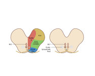 |
Figure 69: Vascular Distribution and Anatomy (Including 4th Nerve) of the Caudal Midbrain | In this axial section of the midbrain at the level of the inferior colliculus, the 4th nuclei are located ventral to the periaqueductal grey, dorsal to the medial longitudinal fasciculus (MLF) and medial to the oculosympathetic tract. Fascicles exit the nucleus dorsally and decussate at the anterior... | Image |
| 132 |
 |
Figure 69: Vascular Distribution and Anatomy (Including 4th Nerve) of the Caudal Midbrain (Supplement) | Image | |
| 133 |
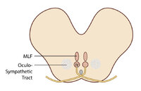 |
Figure 69: Vascular Distribution and Anatomy (Including 4th Nerve) of the Caudal Midbrain (Supplement) | Image | |
| 134 |
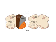 |
Figure 80: Vascular Distribution and Anatomy Relevant to the Medial Medullary Syndrome | This axial section of the medulla highlights those structures that, when damaged, are often responsible for spontaneous upbeat nystagmus (UBN). The nucleus of Roller and nucleus intercalatus normally have an inhibitory influence over the cerebellar flocculus, and when there is a lesion of Roller/int... | Image |
| 135 |
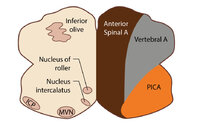 |
Figure 80: Vascular Distribution and Anatomy Relevant to the Medial Medullary Syndrome (Supplement) | Image | |
| 136 |
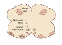 |
Figure 80: Vascular Distribution and Anatomy Relevant to the Medial Medullary Syndrome (Supplement) | Image | |
| 137 |
 |
Five Common Ocular Motor Signs in Cerebellar Disorders - Saccadic Hypermetria, Saccadic Pursuit & VOR Suppression, Gaze-evoked & Rebound Nystagmus | (1) Saccadic hypermetria - an overshoot of the visual target (2) Saccadic smooth pursuit - due to impaired pursuit and low gain, saccades are needed to keep up with the visual target. This gives it a ‘choppy' appearance. (3) Saccadic vestibulo-ocular reflex (VOR) suppression - another... | Image/MovingImage |
| 138 |
 |
Fixation and Gaze Holding | Fixation and gaze-holding: assess for nystagmus or saccadic intrusions by observing the eyes in primary position. Then instruct the patient to look in each position of gaze, and to hold that position to assess for gaze-evoked nystagmus. In doing so, motility can also be evaluated with both eyes view... | Image/MovingImage |
| 139 |
 |
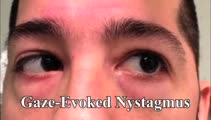
Gaze-Evoked Nystagmus & Slow Saccades Due to Anti-GAD Antibodies in a Patient with Stiff Person Syndrome | This is a 70-year-old woman with a several year long history of imbalance and stiffness. Exam demonstrated axial and lower extremity stiffness, and ocular motor exam demonstrated gaze-evoked nystagmus (e.g., right-beating in right gaze, left-beating in left gaze, up-beating in up gaze), and mild to ... | Image/MovingImage |
| 140 |
|
Gaze-Evoked and Centripetal Nystagmus in Creutzfeldt-Jakob Disease | This is a 65-year-old woman who experienced a progressive cerebellopathy over several months. Initially, she presented with mild gait imbalance and positional vertigo, and there was only apogeotropic positional nystagmus (more pronounced in supine roll test compared to Dix-Hallpike) with a very slig... | Image/MovingImage |
| 141 |
 |
Gaze-Evoked, Rebound, and Centripetal Nystagmus in Cerebellar Degeneration | A 68-year-old female reported a 2-year history of progressive gait imbalance, falls, dizziness and vertical oscillopsia. She described that dizziness and oscillopsia were worst when looking down. There was no family history of ataxia. Composite gaze with fixation was recorded with video-oculography ... | Image/MovingImage |
| 142 |
 |
Gaze-evoked and Rebound Nystagmus in a Cerebellar Syndrome | 𝗢𝗿𝗶𝗴𝗶𝗻𝗮𝗹 𝗗𝗲𝘀𝗰𝗿𝗶𝗽𝘁𝗶𝗼𝗻: 30-yo-man with the subacute onset of a cerebellar syndrome. After extensive evaluation and progression, it was thought that this represented an autoimmune process and there was some improvement with immunosuppression. He ... | Image/MovingImage |
| 143 |
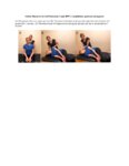 |
Gufoni Maneuver for Left Horizontal Canal BPPV, Canalithiasis (Geotropic Nystagmus) | The Gufoni maneuver may be preferable to the BBQ roll, as the Gufoni maneuver does not require the individual to roll or be in a prone position, making the maneuver more feasible to complete for individuals who are elderly, obese and/or experience immobility. Antecedently, some clinicians remember t... | Text |
| 144 |
 |
Gufoni Maneuver for Left Horizontal Canal BPPV, Canalithiasis (Geotropic Nystagmus) (Video) | The Gufoni maneuver may be preferable to the BBQ roll, as the Gufoni maneuver does not require the individual to roll or be in a prone position, making the maneuver more feasible to complete for individuals who are elderly, obese and/or experience immobility. Antecedently, some clinicians remember t... | Image/MovingImage |
| 145 |
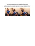 |
Gufoni Maneuver for Right Horizontal Canal-Cupulolithiasis (Apgeotropic Nystagmus) | The Gufoni Maneuver can be used to treat horizontal canal cupulolithaisis. 1. The patient starts in a seated position. 2. The patient transitions quickly to lying on their affected side. 3. The patient lies on their affected side for two minutes with the head in a neutral position. 4. The patient's ... | Text |
| 146 |
 |
Gufoni Maneuver for Right Horizontal Canal-Cupulolithiasis (Apgeotropic Nystagmus) (Video) | The Gufoni Maneuver can be used to treat horizontal canal cupulolithaisis. 1. The patient starts in a seated position. 2. The patient transitions quickly to lying on their affected side. 3. The patient lies on their affected side for two minutes with the head in a neutral position. 4. The patient's ... | Image/MovingImage |
| 147 |
 |
HINTS 'Plus' Patterns in the Acute Vestibular Syndrome Based on Location | HINTS ‘Plus' patterns in the acute vestibular syndrome based on location | Text |
| 148 |
 |
HINTS Exam and Saccadic Dysmetria in Lateral Medullary Stroke | 𝗢𝗿𝗶𝗴𝗶𝗻𝗮𝗹 𝗗𝗲𝘀𝗰𝗿𝗶𝗽𝘁𝗶𝗼𝗻: This is a 50-year-old who experienced the abrupt onset of prolonged vertigo following chiropractic therapy 2 months prior. Initial work-up included an MRI and MR angiogram - MR-diffusion weighted imaging showed an acute l... | Image/MovingImage |
| 149 |
 |
Head Movement Independent ('Sitting') Oscillopsia - A Common Symptom of Nystagmus and Saccadic Intrusions/Oscillations | 𝗢𝗿𝗶𝗴𝗶𝗻𝗮𝗹 𝗗𝗲𝘀𝗰𝗿𝗶𝗽𝘁𝗶𝗼𝗻: This video is an example of what a patient with spontaneous nystagmus or saccadic intrusions/oscillations experiences visually during the abnormal eye movements - i.e., oscillopsia (illusion of movement of the stationary ... | Image/MovingImage |
| 150 |
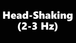 |
Head-Shaking (2-3 Hz) | Head-shaking: instruct the patient to close their eyes and perform active rapid head-shaking at 2-3 Hz for ~15 secs. If a unilateral vestibulopathy is present, head-shaking-induced (contralesional) nystagmus is often provoked, with the slow phase toward the affected ear. With central lesions, the ny... | Image/MovingImage |