AAO-NANOS Neuro-Ophthalmology Clinical Collection: Derived from the AAO-NANOS Clinical Neuro-Ophthalmology collection produced on CD. The images are of selected cases from the NANOS teaching slide exchange, and the CD was produced under the direction of Larry Frohman, MD and Andrew Lee, MD.
The American Academy of Ophthalmology (AAO); The North American Neuro-Ophthalmology Association (NANOS).
NOVEL: https://novel.utah.edu/
TO
Filters: Collection: "ehsl_novel_aao_nanos"
| Title | Creator | Description | ||
|---|---|---|---|---|
| 126 |
 |
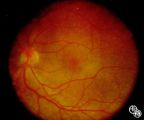
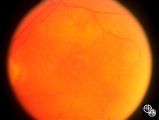
Neuro-Ophthalmic Consequences of Therapy | Mark J. Kupersmith, MD | radiation retinopathy may mimic diabetic or hypertensive optic neuropathy. A history of irradiation to the eye, orbit, or head is mandatory. Radiation retinopathy usually occurs many months after radiation therapy. |
| 127 |
 |
Neuro-Ophthalmic Consequences of Therapy | Mark J. Kupersmith, MD | Radiation causes a vascular retinopathy that may mimic diabetic or hypertensive retinopathy. It does not develop until many months or several years after radiation therapy to the eye, orbit or head. |
| 128 |
 |
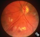
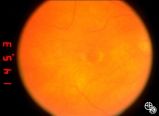
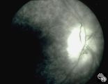
Neuro-Ophthalmic Consequences of Therapy | Joel M. Weinstein, MD | The patient is a 38-year-old woman with oligodendroglioma of the left cerebral hemisphere. The patient received intracarotid BCNU and developed BCNU retinopathy, optic neuropathy, and partial ophthalmoplegia. Image 92_21is from 1 month later and demonstrates hemorrhagic swelling of the disc, retinal... |
| 129 |
 |
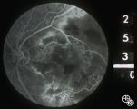
Neuro-Ophthalmic Consequences of Therapy | Joel M. Weinstein, MD | The patient is a 38-year-old woman with oligodendroglioma of the left cerebral hemisphere. The patient received intracarotid BCNU and developed BCNU retinopathy, optic neuropathy, and partial ophthalmoplegia. Image 92_22 Shows late changes, with more severe retinal ischemia, vascular attenuation, an... |
| 130 |
 |
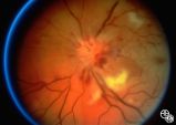
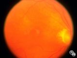
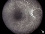
Neuro-Ophthalmic Consequences of Therapy | Larry P. Frohman, MD | This woman presented at age 52, 3 years after radiation therapy for a salivary gland carcinoma extending into the right maxillary sinus. She had received 6000 rads in 30 fractions over 45 days. She presented with 3 weeks of visual loss, with acuity of 20/30, normal color plates, normal fields, and n... |
| 131 |
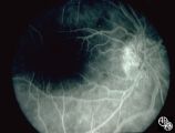 |
Neuro-Ophthalmic Consequences of Therapy | Larry P. Frohman, MD | This woman presented at age 52, 3 years after radiation therapy for a salivary gland carcinoma extending into the right maxillary sinus. She had received 6000 rads in 30 fractions over 45 days. She presented with 3 weeks of visual loss, with acuity of 20/30, normal color plates, normal fields, and n... |
| 132 |
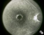 |
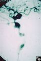
Neuro-Ophthalmic Consequences of Therapy | Don Bienfang, MD | An elderly woman was first seen in 1991, after being on Plaquenil (Hydroxychloroquine Sulfate) for 5 to 6 years, with a new symptomatic disturbance in her reading. Because her blue/yellow color perception was disturbed, and because she had symptoms, the medication was stopped. However the fundi and ... |
| 133 |
 |
Neuro-Ophthalmic Consequences of Therapy | Don Bienfang, MD | An elderly woman was first seen in 1991, after being on Plaquenil (Hydroxychloroquine Sulfate) for 5 to 6 years, with a new symptomatic disturbance in her reading. Because her blue/yellow color perception was disturbed, and because she had symptoms, the medication was stopped. However the fundi and ... |
| 134 |
 |
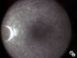
Neuro-Ophthalmic Consequences of Therapy | Don Bienfang, MD | An elderly woman was first seen in 1991, after being on Plaquenil (Hydroxychloroquine Sulfate) for 5 to 6 years, with a new symptomatic disturbance in her reading. Because her blue/yellow color perception was disturbed, and because she had symptoms, the medication was stopped. However the fundi and ... |
| 135 |
 |
Neuro-Ophthalmic Consequences of Therapy | Don Bienfang, MD | An elderly woman was first seen in 1991, after being on Plaquenil (Hydroxychloroquine Sulfate) for 5 to 6 years, with a new symptomatic disturbance in her reading. Because her blue/yellow color perception was disturbed, and because she had symptoms, the medication was stopped. However the fundi and ... |
| 136 |
 |
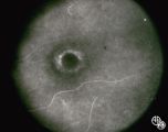
Neuro-Ophthalmic Consequences of Therapy | Don Bienfang, MD | An elderly woman was first seen in 1991, after being on Plaquenil (Hydroxychloroquine Sulfate) for 5 to 6 years, with a new symptomatic disturbance in her reading. Because her blue/yellow color perception was disturbed, and because she had symptoms, the medication was stopped. However the fundi and ... |
| 137 |
 |
Neuro-Ophthalmic Consequences of Therapy | Don Bienfang, MD | An elderly woman was first seen in 1991, after being on Plaquenil (Hydroxychloroquine Sulfate) for 5 to 6 years, with a new symptomatic disturbance in her reading. Because her blue/yellow color perception was disturbed, and because she had symptoms, the medication was stopped. However the fundi and ... |
| 138 |
 |
Neuro-Ophthalmic Consequences of Therapy | Larry P. Frohman, MD | This woman presented at age 52, 3 years after radiation therapy for a salivary gland carcinoma extending into the right maxillary sinus. She had received 6000 rads in 30 fractions over 45 days. She presented with 3 weeks of visual loss, with acuity of 20/30, normal color plates, normal fields, and n... |
| 139 |
 |
Neuro-Ophthalmic Consequences of Therapy | Don Bienfang, MD | An elderly woman was first seen in 1991, after being on Plaquenil (Hydroxychloroquine Sulfate) for 5 to 6 years, with a new symptomatic disturbance in her reading. Because her blue/yellow color perception was disturbed, and because she had symptoms, the medication was stopped. However the fundi and ... |
| 140 |
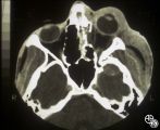 |
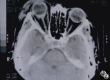
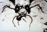
Neuro-Ophthalmic Imaging-CT Scan | Larry P. Frohman, MD | This patient was assaulted with a blunt object and suffered acute blindness due to traumatic optic neuropathy. Note how the lateral orbital wall has been fractured and displaced posteromedially into the region of the anterior optic canal. |
| 141 |
 |
Neuro-Ophthalmic Imaging-CT Scan | Larry P. Frohman, MD | This 39-year-old woman's initial sign was painless, progressive, symmetric ptosis OU, without diurnal variation, that manifested when she was age 17 living in the Dominican Republic. At that time, she had no diplopia or systemic signs. She had no family history of ocular or muscle disease, and no ot... |
| 142 |
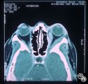 |
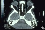
Neuro-Ophthalmic Imaging-CT Scan | Mitchell J. Wolin, MD | Idiopathic orbital pseudotumor is an inflammatory disorder that may effect any part of the ocular anatomy. The site of inflammation determines the nomenclature. For example, involvement of the sclera is referred to as scleritis. And involvement of one or more of the extraocular muscles is referred t... |
| 143 |
 |
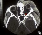
Neuro-Ophthalmic Imaging-CT Scan | Larry P. Frohman, MD | This 70-year-old woman sustained traumatic optic neuropathy in a motor vehicle accident. Note the funnel-shaped hemorrhage within the optic nerve sheath just posterior to the globe. |
| 144 |
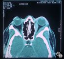 |
Neuro-Ophthalmic Imaging-CT Scan | Mitchell J. Wolin, MD | Idiopathic orbital pseudotumor is an inflammatory disorder that may effect any part of the ocular anatomy. The site of inflammation determines the nomenclature. For example, involvement of the sclera is referred to as scleritis. And involvement of one or more of the extraocular muscles is referred t... |
| 145 |
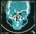 |
Neuro-Ophthalmic Imaging-CT Scan | Mitchell J. Wolin, MD | This is a patient with trauma leading to enucleation, with swelling years later over the implant. This is a presumed chronic abscess between orbit and dura. |
| 146 |
 |
Neuro-Ophthalmic Imaging-CT Scan | Larry P. Frohman, MD | This patient was assaulted with a blunt object and suffered acute blindness due to traumatic optic neuropathy. Note how the lateral orbital wall has been fractured and displaced posteromedially into the region of the anterior optic canal. |
| 147 |
 |
Neuro-Ophthalmic Imaging-Cerebral Angiography | Mark J. Kupersmith, MD | Ehlers-Danlos syndrome is a connective tissue disorder that may affect blood vessels and predispose some affected patients to development of carotid cavernous fistula. Most patients with high-flow direct carotid cavernous sinus fistulas have suffered acute traumatic tears in the internal carotid art... |
| 148 |
 |
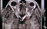
Neuro-Ophthalmic Imaging-MRI | Rosa A. Tang, MD | Aneurisms may result in neuro-ophthalmologic sign and symptoms by direct compression of the afferent or efferent systems or by the secondary effects of hemorrhage. Basilar aneurisms may result in ocular motor deficits such as a unilateral or bilateral third nerve palsy. |
| 149 |
 |
Neuro-Ophthalmic Imaging-MRI | Scott Forman, MD | This 23-year-old right-handed man had a history of idiopathic recurrent optic neuritis. The patient presented with acuity of 20/400 OD and 20/100 OS, with a central scotoma OD and a complete temporal defect OS. MRI with fat suppression and gadolinium revealed enhancement of the intracranial nerve an... |
| 150 |
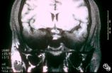 |
Neuro-Ophthalmic Imaging-MRI | Scott Forman, MD | This 23-year-old right-handed man had a history of idiopathic recurrent optic neuritis. The patient presented with acuity of 20/400 OD and 20/100 OS, with a central scotoma OD and a complete temporal defect OS. MRI with fat suppression and gadolinium revealed enhancement of the intracranial nerve an... |