The Health Education Assets Library (HEAL) is a collection of over 22,000 freely available digital materials for health sciences education. The collection is now housed at the University of Utah J. Willard Marriott Digital Library.
TO
Filters: Collection: "ehsl_heal"
| Title | Description | Subject | Collection | ||
|---|---|---|---|---|---|
| 126 |
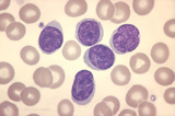 |
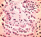
Acute T-cell Leukemia with Multi-cleaved Nuclei | peripheral smear of ATL with multi-cleaved nuclei | Sa0100 | Albert Einstein College of Medicine Gallery of Hematology Images |
| 127 |
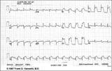 |
Acute anterior MI | Acute anterior MI | Knowledge Weavers ECG | |
| 128 |
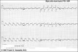 |
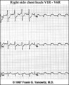
Acute infero-postero-lateral MI | Hyperacute ST segment elevation is seen in leads II, III, aVF (inferior location) and in leads V4-6 (apical lateral wall location). Hyperacute ST depression is seen in leads V1-2 (an expression of posterior wall injury). in addition there are reciprocal ST segment depression changes in leads I an... | Knowledge Weavers ECG | |
| 129 |
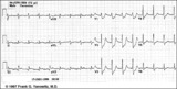 |
Acute inferoposterior MI | Acute inferoposterior MI | Knowledge Weavers ECG | |
| 130 |
 |
Acute postero-lateral MI: precordial leads | Acute postero-lateral MI: precordial leads | Knowledge Weavers ECG | |
| 131 |
 |
Acute pyelonephritis | Acute pyelonephritis | Knowledge Weavers Pathology | |
| 132 |
 |
Acute stasis dermatitis | Acute stasis dermatitis, manifested as redness and oozing. | Knowledge Weavers Dermatology | |
| 133 |
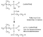 |
Acylation of carnitine by a long chain fatty acyl CoA | Long chain fatty acyl CoA cannot cross the inner mitochondrial membrane to participate in beta-oxidation. The fatty acyl group is therefore transferred to a carrier, carnitine, in a reversible reaction catalyzed by carnitine acyl transferase I. The resulting fatty acyl carnitine crosses the membra... | Knowledge Weavers Fatty Acids | |
| 134 |
 |
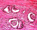
Adenocarcinoma, colon | Adenocarcinoma, colon | Knowledge Weavers Pathology | |
| 135 |
 |
Adenocarcinoma, colon | Adenocarcinoma, colon | Knowledge Weavers Pathology | |
| 136 |
 |
Adenocarcinoma, prostate | Adenocarcinoma, prostate | Knowledge Weavers Pathology | |
| 137 |
 |
Administration of epinephrine into the subcutaneous fat | This demonstrates administration of epinephrine into the subcutaneous fat. | Dosage | Knowledge Weavers Dermatology |
| 138 |
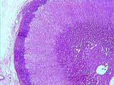 |
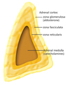
Adrenal | The classical organization of the adrenal gland with a thick capsule and the cortex composed of three layers: the outer zona glomerulosa, the middle zona fasciculata and the inner zona reticularis. Finally, in the center of the gland is the adrenal medulla. UCLA Histology Collection. | Adrenal | UCLA Histology |
| 139 |
 |
Adrenal | This is your best slide of the human adrenal cortex. The capsule is prominent as are the zones of the cortex, the zona glomerulosa, zona fasciculata, and zona reticularis. Deep to the zona reticularis should be the medulla but on this slide, instead of medulla there is a central zone of cortical cel... | Adrenal | UCLA Histology |
| 140 |
 |
Adrenal | Higher magnification of the outer layers of the adrenal cortex including the capsule. The boundary between zona glomerulosa and zona fasciculata is quite irregular. UCLA Histology Collection. | Adrenal | UCLA Histology |
| 141 |
|
Adrenal | The classical organization of the adrenal gland with a thick capsule and the cortex composed of three layers: the outer zona glomerulosa, the middle zona fasciculata and the inner zona reticularis. Finally, in the center of the gland is the adrenal medulla. UCLA Histology Collection. | Adrenal | UCLA Histology |
| 142 |
 |
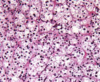
Adrenal - Adrenal Cortex | The layers of adrenal cortex beginning with that next to the capsule are the zona glomerulosa, zona fasciculata, and zona reticularis. The cells of the zona fasciculata look spongy because they contain abundant lipid (steroids) which are dissolved in processing the tissue for histological sectioning... | UCLA Histology | |
| 143 |
 |
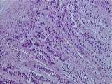
Adrenal - Adrenal Cortex and Adrenal Medulla | This image shows the distinct boundary between the zona reticularis of the cortex and the basophilic chromaffin cells of the medulla. Note that in slide 178, it is difficult to differentiate the layers of the cortex and quite difficult to locate ganglion cells within the medulla. UCLA Histology Coll... | Adrenal | UCLA Histology |
| 144 |
 |
Adrenal - Adrenal Medulla | This high magnification view of the adrenal medulla shows its general lact of organization compared to that of the cortex. A ganglion cell complete with satellite cells is indicated. UCLA Histology Collection. | UCLA Histology | |
| 145 |
 |
Adrenal - Adrenal Medulla | In the center of this image is the atrophied medulla. Note the slight basophilia of the medulla compared ot the eosinophilia of the zona reticularis. UCLA Histology Collection. | adrenal | UCLA Histology |
| 146 |
 |
Adrenal - Adrenal Medulla | In the center of this image is the adrenal medulla. Note its basophilia in contrast to the eosinophilia of the zona reticularis. Compare this image to that of 177-10-1 to appreciate the abnormal atrophy of the medulla in that image. UCLA Histology Collection. | UCLA Histology | |
| 147 |
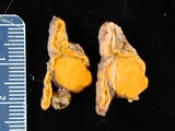 |
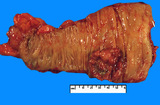
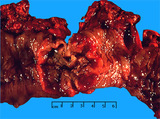
Adrenal Adenoma in Clinical Conn's Syndrome | Adrenal adenoma, aldosterone producing associated with Conn's syndrome. Gross photograph showing 2 contiguous slices of adrenal gland with cortical adenoma. Patient had hyperaldosteronism (Conn's syndrome). | Adrenal Adenoma; Conn Syndrome | HEAL Reviewed Collection |
| 148 |
 |
Adrenal Gland (Labeled) | Adrenal gland. | Royal College of Surgeons in Ireland Illustrations | |
| 149 |
 |
Adrenal adenoma | Adrenal adenoma | Knowledge Weavers Pathology | |
| 150 |
 |
Adrenal cortical adenoma | Adrenal cortical adenoma | Andenoma, Adrenal Cortical | Knowledge Weavers Pathology |