The Health Education Assets Library (HEAL) is a collection of over 22,000 freely available digital materials for health sciences education. The collection is now housed at the University of Utah J. Willard Marriott Digital Library.
TO
Filters: Collection: "ehsl_heal"
| Title | Description | Subject | Collection | ||
|---|---|---|---|---|---|
| 126 |
 |
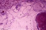
Late cap stage in tooth development - human, embryo | Stain: Azan. From top to bottom: Top side stellate reticulum (enamel pulp) consisting of a network of ectoderm-derived cells; Right side outer dental epithelium with part of the fibrous tooth follicle. This epithelium will further develop downwards as the outer layer of the Hertwig's epithelial root... | oral cavity | Poja Histology Collection - Oral Cavity Subset |
| 127 |
 |
Hilum side of lymph node (human) | Stain: Azan. Site of the hilum of a lymph node includes part of the medulla and medullary cords (1) and medullar sinuses (2). In the hilum (H) dilated efferent lymphatic vessels (3) and draining veins (4) and entering arteries (5) are present. (6) represents adipose tissue embedding the efferent ves... | hilum; lymphatic vessels; medullary cords; paracortex | Poja Histology Collection - Lymphatic Tissues and Organs Subset |
| 128 |
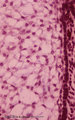 |
Late cap stage in tooth development - human, embryo | Stain: Azan. The stellate reticulum (enamel pulp) consists of a network of ectoderm-derived branched cells and fluid-filled spaces (a.o. proteoglycans). It is a specialized avascular layer as a support and protection for the inner dental epithelial cells. At the right side one layer of cuboidal oute... | oral cavity; tooth development; outer dental epithelium; stellate reticulum | Poja Histology Collection - Oral Cavity Subset |
| 129 |
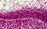 |
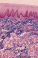
Late cap stage in tooth development - human, embryo | Stain: Azan. From top to bottom: At the top stellate reticulum (enamel pulp) consisting of a loose network of ectoderm-derived cells; Darker stained cell layers of the stratum intermedium; Columnar inner dental epithelium (presecreetory ameloblasts) at the distal side (secretion area) oriented towar... | oral cavity | Poja Histology Collection - Oral Cavity Subset |
| 130 |
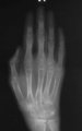 |
Hand X-ray | This radiograph of a child's hand with advanced polyarticular JRA shows subarticular osteopenia and fusion of the metacarpal bones. | Polyarticular Juvenile Rheumatoid Arthritis | HEAL Reviewed Collection |
| 131 |
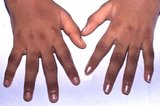 |
Interphalangeal Polyarthritis | In rheumatoid arthritis the proximal interphalangeal joints are more affected than the distal ones. | Polyarticular Juvenile Rheumatoid Arthritis; Interphalangeal Joint | HEAL Reviewed Collection |
| 132 |
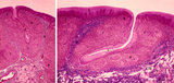 |
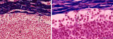
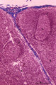
Lingual tonsil ('lymphoepithelial tissue', 'gut-associated lymphatic tissue' or GALT) (human) | Stain: Azan. A: Survey; B: detail of the crypt. The lingual tonsil consists of accumulations of bulging lingual lymphatic follicles in the dorsal part of the tongue behind the terminal sulcus, and belongs to the so-called Waldeyer's ring of pharyngeal lymphatic tissue. The left (A) and right (B) ... | lingual tonsil; GALT; Non-keratinized stratified squamous epithelium; crypt | Poja Histology Collection - Lymphatic Tissues and Organs Subset |
| 133 |
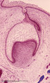 |
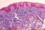
Late cap stage of tooth development - human, embryo; low magnification | Stain: Azan. From top to bottom: Stratified ectoderm with a distinct basal layer (red line) of cuboid cells; Dental lamina giving rise to the cap stage (center) and to the primordium of permanent tooth (right); Odontogenic organ or enamel organ (future deciduous tooth surrounded by fibrous tooth fol... | oral cavity; dental lamina | Poja Histology Collection - Oral Cavity Subset |
| 134 |
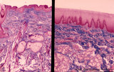 |
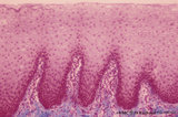
Lip (human), outer (left) and inner side (right) | Stain: Azan. Left image: keratinized squamous epithelium with thin red cornified layer, hair follicle, sebaceous glands and skeletal muscle fibers (orbicularis oris). Right image: non-keratinized epithelium with high, narrow dermal papillae and more capillaries. Mixed labial glands (seromucous), f... | oral cavity; lining mucosa | Poja Histology Collection - Oral Cavity Subset |
| 135 |
 |
Lip (human), region between red zone (vermilion border) and mucosa inner surface | Stain: Azan. Bundles of skeletal muscle fibers (musculus orbicularis oris), highly vascularized lamina propria. Note the narrow dermal papillae on the left side (inner lip) and the broad irregular papillae on the right side (red zone of the lip). | oral cavity; lining mucosa; red zone; vermilion border | Poja Histology Collection - Oral Cavity Subset |
| 136 |
 |
Lip (human), mucous inner surface | Stain: Azan. Non-keratinized epithelium with high, narrow dermal papillae and many capillaries. Mixed labial glands (seromucous) and few skeletal muscle fibers (orbicularis oris) in the submucosa. | oral cavity; lining mucosa; labial glands | Poja Histology Collection - Oral Cavity Subset |
| 137 |
 |
Lip (human), mucoserous labial glands in mucous inner surface | Stain: Azan. Mixed labial glands with serous demilunes (von Ebner-Giannuzzi), a few myoepithelial cells, and a striated duct (right upper corner). | oral cavity; lining mucosa; labial glands | Poja Histology Collection - Oral Cavity Subset |
| 138 |
 |
Lip (human), mucous inner surface | Stain: Azan. Non-keratinized epithelium with high, narrow dermal papillae and many capillaries. Note lymphocytic infiltrates and the capillaries in the dermal papillae. | oral cavity; lining mucosa | Poja Histology Collection - Oral Cavity Subset |
| 139 |
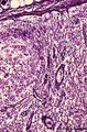 |
Lymph node (human) | Stain: Silver stain (Gomori). Due to the argyrophilia of reticular fibres the reticular network is black-stained among others such as collagen type I, III, IV. From the fibrous capsule (1) a meshwork of fine reticular fibres penetrates into the cortex of a lymph node crossing the subcapsular (or m... | germinal center; argyrophilia; reticular fibers | Poja Histology Collection - Lymphatic Tissues and Organs Subset |
| 140 |
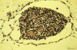 |
Lymph node (fetus, human) | Stain: Trichrome (Goldner). Along the course of lymphatic vessels lymph nodes develop. Lymphatic vessels dilate and form lymphatic sacs and lymphocytes aggregate around these sacs. A tiny developing lymph node with dense-stained thymocyte nuclei is closely associated with dilated lymph capillaries. ... | fetal lymph node; development | Poja Histology Collection - Lymphatic Tissues and Organs Subset |
| 141 |
 |
Lymph node (human) | Stain: Azan. Left: subcapsular (or marginal) sinus in lymph node. Right: higher magnification. Both pictures show a thick capsule (1), at (2) subcapsular (or marginal) sinus filled with lymphocytes indicate (lining) littoral cells (?). Reticular cells (3) are localized perpendicularly through sin... | subcapsular sinus; reticular cell | Poja Histology Collection - Lymphatic Tissues and Organs Subset |
| 142 |
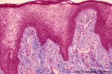 |
Lip (human), transitional zone (red zone or vermilion border) | Stain: Azan. Slightly cornified epithelium with high irregular dermal papillae and many capillaries. Note the epithelium is thicker, but less cornified than the epidermis. The red color of the lips is due to the rich vascularity of the lamina propria and the lucidity of the epithelium. | oral cavity; lining mucosa; red zone; vermilion border | Poja Histology Collection - Oral Cavity Subset |
| 143 |
 |
Lip (human), transitional zone (red zone or vermilion border) | Stain: Azan. Slightly cornified epithelium with high irregular dermal papillae and many capillaries. Fat cells and skeletal muscle cells are located in the submucosa. | oral cavity; lining mucosa; red zone; vermilion border | Poja Histology Collection - Oral Cavity Subset |
| 144 |
 |
Lymph Nodes of Head and Neck (Labeled) | Lymph nodes. Head. Neck. | Submental; Submandibular; Deep Cervical; Jugulodigastric; Mastoid; Occipital; Parotid | Royal College of Surgeons in Ireland Illustrations |
| 145 |
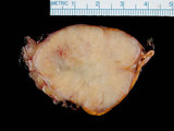 |
Lung carcinoma metastatic to adrenal gland | This is a gross photograph of a non-small cell carcinoma metastatic to the adrenal gland. | HEAL Reviewed Collection | |
| 146 |
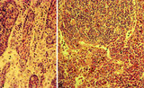 |
Lymph nodes (rat) | Stain: Hematoxylin & pyronin. Pyronin was formerly used to demonstrate young blast cell types rich in polysomes and packed rough endoplasmic reticulum (RER) resulting in a reddish stain of the cytoplasm. A: Part of the medulla with medullary cords (1) and medullary sinuses (2) close to the hilum o... | infection; medullar cord; plasma cells ; macrophages | Poja Histology Collection - Lymphatic Tissues and Organs Subset |
| 147 |
 |
Lymph node (human) | Stain: Azan. Part of the cortex of a lymph node with the capsule (1) and subcapsular (or marginal) sinus (2) filled with lymphocytes. From the blue-stained dense capsule a long trabecula (3) flanked by paratrabecular (intermediate) sinuses penetrates between the paracortical areas (6). Paratrabecula... | Poja Histology Collection - Lymphatic Tissues and Organs Subset | |
| 148 |
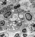 |
Lymph node (rat) | Electron microscopy. In the paracortical area (inner cortex) of a lymph node interdigitating cells (1) are found (so-called IDC, antigen-presenting cell or APC). They show branched extensions (3) in between the closely apposed surrounding T lymphocytes (2). In the cytoplasm of the IDC lysosomal inc... | electron microscopy; interdigitating cell; antigen-presenting cell | Poja Histology Collection - Lymphatic Tissues and Organs Subset |
| 149 |
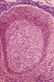 |
Lymph node (human) | Stain: Azan. Part of the cortex of a lymph node with the capsule (1) and subcapsular (or marginal) sinus (2) filled with lymphocytes. Below the subcapsular sinus a darkly stained rim or corona (3) of small lymphocytes (B cells) surrounds the lucid stained germinal centre (4). The white spaces repre... | follicle; germinal center | Poja Histology Collection - Lymphatic Tissues and Organs Subset |
| 150 |
 |
Lymph node (mouse) | Stain: Hematoxylin & eosin. For comparison with the human lymph node a survey of a small rodent lymph node is demonstrated. Basically the same divisions are found: (1) a cortex (outer cortex zone, nodular cortex or superficial cortex) covered with a (2) thin capsule and subcapsular sinus; a paracor... | follicle; hilum; medulla; cortex | Poja Histology Collection - Lymphatic Tissues and Organs Subset |