The Health Education Assets Library (HEAL) is a collection of over 22,000 freely available digital materials for health sciences education. The collection is now housed at the University of Utah J. Willard Marriott Digital Library.
TO
| Title | Description | Subject | Collection | ||
|---|---|---|---|---|---|
| 126 |
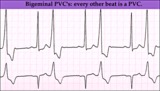 |
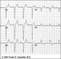
Ventricular bigeminy - marquette | Ventricular bigeminy - marquette | Knowledge Weavers ECG | |
| 127 |
 |
LBBB and 2nd degree AV block, mobitz type I | Mobitz II 2nd degree AV block is usually a sign of bilateral bundle branch disease. One of the two bundle branches should be completely blocked; in this example the left bundle is blocked. The nonconducted sinus P waves are most likely blocked in the right bundle which exhibits 2nd degree block. ... | Knowledge Weavers ECG | |
| 128 |
 |
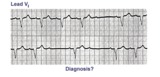
Infero-posterior MI&RBBB | Deep Q waves in II, III, aVF plus tall R waves in V1-2 are evidence for this infero-posterior MI. The wide QRS (>0.12s) and RR' complex in V1 are evidence for RBBB. Any time RBBB has an initial R in V1 equal to or greater than the R', true posterior MI must be considered. Q waves in V5-6 suggest a... | Knowledge Weavers ECG | |
| 129 |
 |
Extensive anterior/anterolateral MI: recent | Significant pathologic Q-waves (V2-6, I, aVL) plus marked ST segment elevation are evidence for this large anterior/anterolateral MI. The exact age of the infarction cannot be determined without clinical correlation and previous ECGs, but this is likely a recent MI. | Knowledge Weavers ECG | |
| 130 |
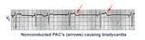 |
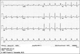
Nonconducted PAC's slowing the heart rate | Consecutive nonconducted PAC's, indicated by arrows, can significantly slow the heart rate. Note the distortion of the ST-T waves caused by the PAC. A hint in recognizing nonconducted PAC's is to find conducted PAC's in the same rhythm strip. | Knowledge Weavers ECG | |
| 131 |
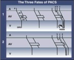 |
three fates of PAC's: 1. normal conduction; 2. aberrant conduction; 3. non-conduction | three fates of PAC's: 1. normal conduction; 2. aberrant conduction; 3. non-conduction | Knowledge Weavers ECG | |
| 132 |
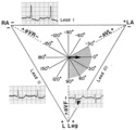 |
QRS axis = 0 degrees | Lead aVF is isoelectric; lead I is positive; therefore, the QRS axis is 0 degrees. | Knowledge Weavers ECG | |
| 133 |
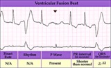 |
Ventricular fusion beat - marquette | Ventricular fusion beat - marquette | Knowledge Weavers ECG | |
| 134 |
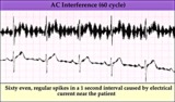 |
60 cycle artifact - marquette | 60 cycle artifact - marquette | Knowledge Weavers ECG | |
| 135 |
 |
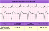
LBBB: precordial leads | LBBB: precordial leads | Knowledge Weavers ECG | |
| 136 |
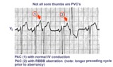 |
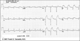
Not all sore thumbs are ventricular in origin | PACs have three fates: normal conduction into ventricles, aberrant conduction in ventricles due to bundle branch or fascicular block, and non-conduction due to block in AV junction. In this example PAC 1 is normally conducted and PAC 2 is conducted with RBBB aberration. The longer preceding cycle ... | Knowledge Weavers ECG | |
| 137 |
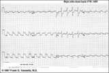 |
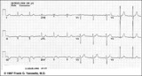
Acute infero-postero-lateral MI | Hyperacute ST segment elevation is seen in leads II, III, aVF (inferior location) and in leads V4-6 (apical lateral wall location). Hyperacute ST depression is seen in leads V1-2 (an expression of posterior wall injury). in addition there are reciprocal ST segment depression changes in leads I an... | Knowledge Weavers ECG | |
| 138 |
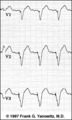 |
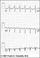
Ventricular Pacemaker Rhythm: V1-3 | Note the small pacemaker spikes before the QRS complexes. In addition, the QRS complex in V1-3 exhibits ventricular ectopic morphology; i.e., there is a slur or notch at the beginning of the S wave, and >60ms delay from onset to QRS to nadir of S wave. This rules against a supraventricular rhythm wi... | Knowledge Weavers ECG | |
| 139 |
 |
Inferior MI: fully evolved | Significant pathologic Q-waves are seen in leads II, III, aVF along with resolving ST segment elevation and symmetrical T wave inversion. This is a classic inferior MI. | Knowledge Weavers ECG | |
| 140 |
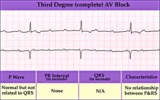 |
Complete AV block (3rd degree) with junctional rhythm | Complete AV block (3rd degree) with junctional rhythm | Knowledge Weavers ECG | |
| 141 |
 |
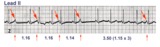
QRS axis = +90 degrees | Lead I is isoelectric; II and III are positive; the axis is +90 degrees. | Knowledge Weavers ECG | |
| 142 |
 |
Left bundle branch block - marquette | Left bundle branch block - marquette | Knowledge Weavers ECG | |
| 143 |
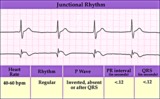 |
Junctional escape rhythm | Junctional escape rhythm | Knowledge Weavers ECG | |
| 144 |
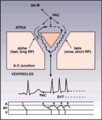 |
Diagram: AV nodal reentrant tachycardia | The AV node often has dual pathways; in this diagram the alpha pathway is fast, but has a long refractory period; the beta pathway is conducts more slowly, but recovers faster.In sinus rhythm the faster alpha pathway is used and accounts for the normal PR interval. When a PAC occurs, however, the i... | Knowledge Weavers ECG | |
| 145 |
 |
Atrial parasystole | In atrial parasystole non-fixed coupled PACs, shown by arrows, occur at a common inter-ectopic interval or at multiples of this interval. Atrial fusions, not shown here, may also occur when the PAC occurs in close temporal proximity to the sinus impulse. | Knowledge Weavers ECG | |
| 146 |
 |
Atrial flutter with 2:1 conduction: leads II, III, V1 | Atrial flutter with 2:1 conduction: leads II, III, V1 | Knowledge Weavers ECG | |
| 147 |
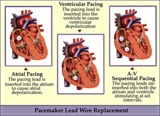 |
Pacemaker lead wire placement diagram - marquette | Pacemaker lead wire placement diagram - marquette | Knowledge Weavers ECG | |
| 148 |
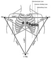 |
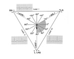
Frontal and horizontal plane lead diagram | Frontal and horizontal plane lead diagram | Knowledge Weavers ECG | |
| 149 |
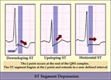 |
ST segment diagram - marquette | ST segment diagram - marquette | Knowledge Weavers ECG | |
| 150 |
 |
Atrial bigeminy - marquette | Atrial bigeminy - marquette | Knowledge Weavers ECG |