AAO-NANOS Neuro-Ophthalmology Clinical Collection: Derived from the AAO-NANOS Clinical Neuro-Ophthalmology collection produced on CD. The images are of selected cases from the NANOS teaching slide exchange, and the CD was produced under the direction of Larry Frohman, MD and Andrew Lee, MD.
The American Academy of Ophthalmology (AAO); The North American Neuro-Ophthalmology Association (NANOS).
NOVEL: https://novel.utah.edu/
TO
| Title | Creator | Description | ||
|---|---|---|---|---|
| 126 |
 |
Isolated Optic Neuritis/Neuropathy | Larry P. Frohman, MD | The patient is a 62-year-old female who presented in August 1996 with visual loss OD that she first noted as loss of her superior field in May 1996. She felt that it had been static since, and perhaps was even a little better in the week before she was seen. There was no pain, even with ocular rotat... |
| 127 |
 |
Systemic Disorders With Optic Nerve and Retinal Findings | Larry P. Frohman, MD | A 42-year old woman presented with a history of severe brow pain and 4 days of progressive visual loss OD. There was no increased pain on ocular rotation. Aside from heavy menses, she denied any significant past medical history. Her examination revealed acuity NLP OD, 20/25 OS; color vision 9/10 OS;... |
| 128 |
 |
Neuro-Ophthalmic Vascular Disease | Steven A. Newman, MD | Occlusion of a branch or central retinal artery may result in acute visual loss. The ophthalmoscopic findings are retinal whitening due to ischemic retina in the distribution of the occluded artery. Sparing or selective involvement of cilioretinal artery branches may occur. Patients with a central r... |
| 129 |
 |
Systemic Disorders With Optic Nerve and Retinal Findings | John A. Charley, MD | A hypercoagulable state may result from deficiency of various factors involved in the clotting pathway, such as factor V. This may result in venous thrombosis and a branch or central retinal vein occlusion. Consideration for testing for an underlying coagulopathy, including the factor V deficiency, ... |
| 130 |
 |
Systemic Disorders With Optic Nerve and Retinal Findings | John A. Charley, MD | A hypercoagulable state may result from deficiency of various factors involved in the clotting pathway, such as factor V. This may result in venous thrombosis and a branch or central retinal vein occlusion. Consideration for testing for an underlying coagulopathy, including the factor V deficiency, ... |
| 131 |
 |
Orbital Tumors | Larry P. Frohman, MD | This 30-year-old man had a retrobulbar intraconal mass OS. The CT scans showed a heterogeneous lobulated enhancing mass, 2.2 x 1.9 x 1.8 cm. The case beautifully exhibits chorodial folds. The ultrasound showed internal reflectivity. The patient refused surgery. Pair with Images 97_60, 97_62, 97_63, ... |
| 132 |
 |
Orbital Tumors | Larry P. Frohman, MD | This 30-year-old man had a retrobulbar intraconal mass OS. The CT scans showed a heterogeneous lobulated enhancing mass, 2.2 x 1.9 x 1.8 cm. The case beautifully exhibits chorodial folds. The ultrasound showed internal reflectivity. The patient refused surgery. Pair with Images 97_60, 97_61, 97_63, ... |
| 133 |
 |
Orbital Tumors | Larry P. Frohman, MD | This 30-year-old man had a retrobulbar intraconal mass OS. The CT scans showed a heterogeneous lobulated enhancing mass, 2.2 x 1.9 x 1.8 cm. The case beautifully exhibits chorodial folds. The ultrasound showed internal reflectivity. The patient refused surgery. Pair with Images 97_61, 97_62, 97_63, ... |
| 134 |
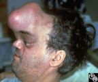 |
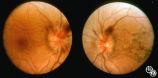
Neuro-Ophthalmic Manifestations of Brain Tumors | Steven Galetta, MD | The patient is a 45-year-old recluse found to harbor this frontal lobe mass. Remarkably, this patient had only mild bilateral optic neuropathies with visual acuities in the 20/25 range. This right disc was mildly swollen and the left mildly pale. He could not fit into the fundus camera for disc phot... |
| 135 |
 |
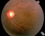
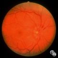
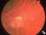
Isolated Congenital Optic Disc Anomalies | Larry P. Frohman, MD | This 63-year-old man with amblyopia OD was seen for a question of ischemic optic neuropathy with a pale, swollen disc OD. The correct diagnosis is an exophytic capillary angioma of the optic nerve head. Disease/Diagnosis: Capillary Angioma. |
| 136 |
 |
Optic Neuropathies | Larry P. Frohman, MD | This healthy 29-year-old man with dense amblyopia OS presented with a foreign-body sensation OS and further visual loss in his amblyopic eye. He was noted to have bilateral disc edema and lesions in the left eye consistent with unilateral acute multifocal placoid pigment epitheliopathy (AMPPE). He r... |
| 137 |
 |
Optic Neuropathies | Larry P. Frohman, MD | This healthy 29-year-old man with dense amblyopia OS presented with a foreign-body sensation OS and further visual loss in his amblyopic eye. He was noted to have bilateral disc edema and lesions in the left eye consistent with unilateral acute multifocal placoid pigment epitheliopathy (AMPPE). He r... |
| 138 |
 |
Optic Neuropathies | Larry P. Frohman, MD | This healthy 29-year-old man with dense amblyopia OS presented with a foreign-body sensation OS and further visual loss in his amblyopic eye. He was noted to have bilateral disc edema and lesions in the left eye consistent with unilateral acute multifocal placoid pigment epitheliopathy (AMPPE). He r... |
| 139 |
 |
Optic Neuropathies | Larry P. Frohman, MD | This healthy 29-year-old man with dense amblyopia OS presented with a foreign-body sensation OS and further visual loss in his amblyopic eye. He was noted to have bilateral disc edema and lesions in the left eye consistent with unilateral acute multifocal placoid pigment epitheliopathy (AMPPE). He r... |
| 140 |
 |
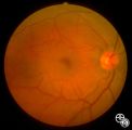
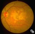
Systemic Disorders With Optic Nerve and Retinal Findings | Larry P. Frohman, MD | At age 41, in 1984, this woman, who grew up in the Ohio River Valley, had 3 days of ocular pain OD, and her vision declined to 20/80 OD she has had no visual changes since, nor has she had any other neurologic symptoms. The ""presumed"" diagnosis is optic neuropathy in presumed ocular histoplasmosis... |
| 141 |
 |
Systemic Disorders With Optic Nerve and Retinal Findings | Larry P. Frohman, MD | This 57-year-old man had a neuro-ophthalmology consult, requested the night before his 2-cm pituitary tumor was to be resected. His examination revealed his acuities to be 20/70 OU, with a visual field not consistent with chiasmal compression. The fundus appearance, with peripheral salt and pepperin... |
| 142 |
 |
Neuro-Ophthalmic Vascular Disease | Steven A. Newman, MD | Occlusion of a branch or central retinal artery may result in acute visual loss. The ophthalmoscopic findings are retinal whitening due to ischemic retina in the distribution of the occluded artery. Sparing or selective involvement of cilioretinal artery branches may occur. Patients with a central r... |
| 143 |
 |
Neuro-Ophthalmic Vascular Disease | Steven A. Newman, MD | Occlusion of a branch or central retinal artery may result in acute visual loss. The ophthalmoscopic findings are retinal whitening due to ischemic retina in the distribution of the occluded artery. Sparing or selective involvement of cilioretinal artery branches may occur. Patients with a central r... |
| 144 |
 |
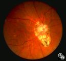
Systemic Disorders With Optic Nerve and Retinal Findings | Larry P. Frohman, MD | This is a 32-year-old HIV-positive man with anterior uveitis, vitritis, and bilateral papillitis from syphilis. With intravenous penicillin treatment, the optic discs and vision returned to normal. |
| 145 |
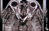 |
Neuro-Ophthalmic Imaging-MRI | Scott Forman, MD | This 23-year-old right-handed man had a history of idiopathic recurrent optic neuritis. The patient presented with acuity of 20/400 OD and 20/100 OS, with a central scotoma OD and a complete temporal defect OS. MRI with fat suppression and gadolinium revealed enhancement of the intracranial nerve an... |
| 146 |
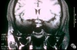 |
Neuro-Ophthalmic Imaging-MRI | Scott Forman, MD | This 23-year-old right-handed man had a history of idiopathic recurrent optic neuritis. The patient presented with acuity of 20/400 OD and 20/100 OS, with a central scotoma OD and a complete temporal defect OS. MRI with fat suppression and gadolinium revealed enhancement of the intracranial nerve an... |
| 147 |
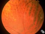 |
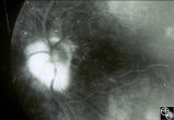
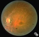
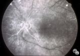
Systemic Disorders With Optic Nerve and Retinal Findings | Robert L. Lesser, MD | Intraocular lymphoma may present with an unexplained vitritis, optic disc infiltration, or choroidal infiltration. One unusual manifestation of large-cell lymphoma is this leopard-spot appearance. Pair with 94_32, 94_33, and 94_35. This is a fundus photo. |
| 148 |
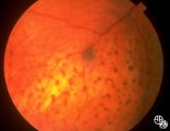 |
Systemic Disorders With Optic Nerve and Retinal Findings | Robert L. Lesser, MD | Intraocular lymphoma may present with an unexplained vitritis, optic disc infiltration, or choroidal infiltration. One unusual manifestation of large-cell lymphoma is this leopard-spot appearance. Pair with 94_32, 94_34, and 94_35. This is a fundus photo. |
| 149 |
 |
Systemic Disorders With Optic Nerve and Retinal Findings | Robert L. Lesser, MD | Intraocular lymphoma may present with an unexplained vitritis, optic disc infiltration, or choroidal infiltration. One unusual manifestation of large-cell lymphoma is this leopard-spot appearance. Pair with 94_33, 94_34, and 94_35. This is a fundus photo. |
| 150 |
 |
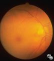
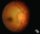
Optic Neuropathies | Daniel M. Jacobson MD | This 28-year-old otherwise-healthy woman was referred to for treatment of what was thought to be optic neuritis OD. Three weeks earlier she had noted a dark inferior scotoma OD that progressed to involve fixation over the next 10-12 days. She experienced photopsias OD at the onset. She had no viral ... |