The Health Education Assets Library (HEAL) is a collection of over 22,000 freely available digital materials for health sciences education. The collection is now housed at the University of Utah J. Willard Marriott Digital Library.
TO
| Title | Description | Subject | Collection | ||
|---|---|---|---|---|---|
| 126 |
 |
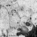
Pulpo-dentinal complex in longitudinal section of tooth - secondary dentin, human, adult | Stain: Hematoxylin and eosin. From left to right: - dentinal tubules as fine striation in dentin; - vertical course of incremental lines (von Ebner); - a darker stained broad zone of secondary dentin; - pulp chamber with crowding of odontoblasts close to secondary, newly formed dentin; - connective ... | oral cavity; secondary dentin | Poja Histology Collection - Oral Cavity Subset |
| 127 |
 |
Pulp canal of tooth - longitudinal section; human, adult | Stain: Hematoxylin and eosin. Centrally three thick nerve bundles. In between blood vessel filled with erythrocytes. At the right side longitudinal course of slender thin-walled blood vessels (arterioles) which pass into thin-walled capillaries with a wide lumen. | oral cavity; pulp canal | Poja Histology Collection - Oral Cavity Subset |
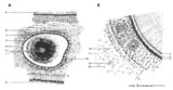
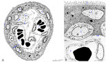
| 128 |
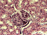 |
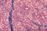
Renal corpuscle | This section of renal corpuscle clearly shows the glomerulus with vascular and urinary poles. Adjacent to the vascular pole is the macula densa region of the distal tubule. At the urinary pole, the proximal convoluted tubule drains the urinary space. | Renal Corpuscle | HEAL Reviewed Collection |
| 129 |
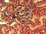 |
Renal corpuscle | The section of renal corpuscle includes the parietal layer of the renal capsule, urinary space and glomerulus. The vascular pole and macula densa region of the distal tubule are represented. | Renal Corpuscle | HEAL Reviewed Collection |
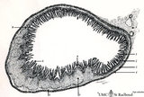
| 130 |
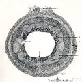 |
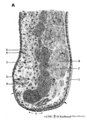
Scheme of appendix ('gut-associated lymphatic tissue' or GALT) (human) | Survey vermiform appendix (see also Digestive System: Appendix) A large amount of non-encapsulated diffuse lymphatic tissue or mucosa-associated lymphatic tissue (MALT) is located in the subepithelial lamina propria (e.g. respiratory passages, genitourinary tract). The gut-associated lymphatic tis... | GALT; follicle; scheme; germinal center | Poja Histology Collection - Lymphatic Tissues and Organs Subset |
| 131 |
 |
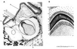
Scheme of cross sectional tooth in alveolar bone - cat | A. Survey of tooth (in alveolar bone) magnification x 30 objective. B. Peripheral part of tooth (in alveolar bone) magnification x 300 objective. 1. radicular pulp; 2. odontoblast layer; 3. dentin (radial arranged dentinal tubules); 4. Tomes' granular layer; 5. cementum; 6. fibers of period... | oral cavity; alveolar bone | Poja Histology Collection - Oral Cavity Subset |
| 132 |
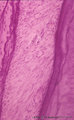 |
Root of tooth within osseous socket - cross-section; human, adult | Stain: Hematoxylin and eosin. From left to right: bundle bone of alveolus; oblique fibers of periodontal ligament with blood vessels (broad layer); flattened cementoblasts settled on a thin light rim of cementoid; darker stained acellular cementum, the so-called acellular extrinsic fiber cementum a... | oral cavity; acellular cementum | Poja Histology Collection - Oral Cavity Subset |
| 133 |
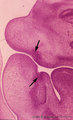 |
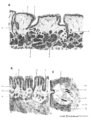
Sagittal section through tooth buds in jaws during tooth development - human, embryo, 6 weeks; low magnification | Stain: hematoxylin and eosin. At the top the upper jaw (maxilla) opposite the lower jaw (mandible). At the right side part of the tongue in the oral cavity. Arrows point to epithelial proliferations (future tooth bud formation) that are surrounded by condensation of mesenchymal cells (neural crest m... | oral cavity; tooth bud; tooth development | Poja Histology Collection - Oral Cavity Subset |
| 134 |
 |
Scheme of compound glands in the oral cavity - human, adult | A. parotid gland (serous gland) magnification x 350 objective. B. sublingual gland (mucous part) magnification x 350 objective. C. sublingual gland (mixed part) magnification x 350 objective. D. simplified scheme of compound serous/mucous gland. 1. myoepithelial cell; 2. serous acinus; 3. stri... | oral cavity | Poja Histology Collection - Oral Cavity Subset |
| 135 |
 |
Scheme of circumvallate papillae of the tongue (human, adult) | 1. (circum)vallate papilla; 2. non-keratinized stratified squamous epithelium; 3. taste bud; 4. serous gland (von Ebner); 5. draining duct (of serous gland); 6. lingual muscle | oral cavity; von Ebner; circumvallate papillae | Poja Histology Collection - Oral Cavity Subset |
| 136 |
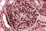 |
Renal corpuscle | This section of renal corpuscle shows the glomerulus with vascular pole and macula densa region of the distal tubule. At the urinary pole, the proximal convoluted tubule can be seen draining the urinary space. | Renal Corpuscle | HEAL Reviewed Collection |
| 137 |
 |
Scheme of lingual tonsil ('lymphoepithelial tissue') | Lingual tonsil (consisting of the accumulation of folliculi linguales). The root of the tongue contains invaginations or crypts or narrow caverns (3). In these crypts the ducts of the mucous glands (8) end up. The crypts are lined by multilayered, non-keratinizing squamous epithelium (2) and are sur... | lingual tonsil; scheme; germinal center; follicle | Poja Histology Collection - Lymphatic Tissues and Organs Subset |
| 138 |
 |
Scheme of ileum with Peyers patches (gut-associated lymphatic tissue or GALT) (dog) | Survey ileum (see also Digestive System: Ileum) A large amount of non-encapsulated diffuse lymphatic tissue or mucosa-associated lymphatic tissue (MALT) is located in the subepithelial lamina propria (e.g. respiratory passages, genitourinary tract). The gut-associated lymphatic tissues (GALT) are ... | GALT; follicle; scheme; germinal center | Poja Histology Collection - Lymphatic Tissues and Organs Subset |
| 139 |
 |
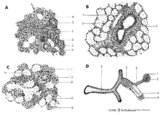
Scheme of electron microscopy of tertiary villus (human placenta, midpregnancy) | (See also POJA-L1227). (A) Survey and (B) detail of a tertiary placental villus lined by the multinucleated syncytiotrophoblast cell (1) (STC). The apex shows protrusions and extensive microvilli of varying sizes (brushborder, 2). Nuclei are sectioned at different levels and the cytoplasm contains a... | placenta; tertiary villi; placental barrier; electron microscopy | Poja Histology Collection - Placenta |
| 140 |
 |
Scheme of palatine and pharyngeal tonsils ('lymphoepithelial tissues') (human) | The palatine tonsils are covered by the multilayered, non-keratinizing squamous epithelium of the oral cavity (A-1). In contrast to the palatine and lingual tonsils, the epipharyngeal tonsil has a multilayered ciliated epithelium (respiratory-like epithelium, B-9). The Waldeyer's tonsillar ring i... | germinal center; scheme; follicle | Poja Histology Collection - Lymphatic Tissues and Organs Subset |
| 141 |
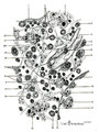 |
Scheme of reticular tissue in lymph node | Lymph node stroma does not contain stromal cells but instead the reticular meshwork is build by reticular cell types, macrophages and lymphoid cells and myeloid cells. Route (A) in the scheme represents morphological differentiation path from B lymphocytes to plasma cells. 1 endothelial ce... | reticular tissue; scheme; follicle; germinal center | Poja Histology Collection - Lymphatic Tissues and Organs Subset |
| 142 |
 |
Scheme of tongue papillae and foliate papillae | A. Circumvallate papillae (tongue, human, adult) magnification x 35 objective. B. Foliate papillae (tongue, rabbit) magnification x 85 objective. C. Taste bud in foliate papilla (tongue, rabbit) magnification x 750 objective. 1. (circum)vallate papilla; 2. non-keratinized stratified squamous epit... | oral cavity; von Ebner; circumvallate papillae; foliate papillae | Poja Histology Collection - Oral Cavity Subset |
| 143 |
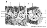 |
Scheme of tongue papillae and taste buds | B. Scheme of foliate papillae of the tongue (rabbit) magnification x 85 objective. C. Scheme of taste bud in foliate papilla of the tongue (rabbit). 2. non-keratinized stratified squamous epithelium; 3. taste bud; 4. serous gland (von Ebner); 5. draining duct (of serous gland); 7. foliate papil... | oral cavity; von Ebner; foliate papillae | Poja Histology Collection - Oral Cavity Subset |
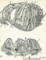
| 144 |
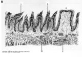 |
Scheme of filiform and fungiform papillae of the tongue - human, adult | 5. filiform papillae; 6. fungiform papillae; 8. locally cornification on top of filiform papillae; 9. lingual muscle; 10. lingual glands | oral cavity; papillae | Poja Histology Collection - Oral Cavity Subset |
| 145 |
 |
Section of lymph node (human) | Stain: Silver stain (Gomori). Due to the argyrophilia the reticular fibers are black-stained. They are derived from the fibrous capsule and penetrate into the deep cortex (i.e. paracortical area) and are embedded among other fibers such as collagen type III, IV. Note the postcapillary venules (*) ... | high endothelial venule (HEV); paracortex; argyrophilia; paracortex | Poja Histology Collection - Lymphatic Tissues and Organs Subset |
| 146 |
 |
Secretory ameloblasts in tooth development - bell stage, gerbil, postnatal | Electronmicroscopy. Cross-sectioned distal sides of ameloblasts (secretion areas or Tomes' processes) with numerous organelles. The dark stained vesicles represent secretory granules contain among others amelogenin. At the right bottom corner intercellular spaces are filled with dark-stained organi... | oral cavity; enamel prisms | Poja Histology Collection - Oral Cavity Subset |
| 147 |
 |
Scheme of tooth development (human) | A. Survey of tooth germ (bell stage, embryo) magnification x 35 objective. B. Formation of enamel and dentin (bell stage, embryo) magnification x 350 objective. 1. pulp organ; 2. odontoblasts; 3. mantle predentin; 4. dentin; 5. enamel; 6. initial enamel; 7. inner dental epithelium (ameloblasts);... | oral cavity; dental lamina; bell stage | Poja Histology Collection - Oral Cavity Subset |
| 148 |
 |
Spiral artery in endometrium (human, midpregnancy) | Stain: Hematoxylin-eosin. (A) cross-sections of spiral arteries in endometrium. (B) semi-polarized section of part of a spiral artery. Dark-stained large trophoblast cells (3), partly replacing the cuboidal hypertrophic (B2) endothelial cells. (4) smooth muscle cells. (L) lumen of vessel with cell... | placenta; spiral artery; endometrium; trophoblast | Poja Histology Collection - Placenta |
| 149 |
 |
Scheme survey lip - human, adult | 1. red (transitional) zone; 2. keratinized stratified squamous epithelium (outer side); 3. non-keratinized stratified squamous epithelium (inner side); 4. hair shaft; 5. hair follicle; 6. sebaceous gland; 7. cross-section of circumoral muscle; 8. labial glands with draining ducts; ... | oral cavity | Poja Histology Collection - Oral Cavity Subset |
| 150 |
 |
Sublingual gland (human) | Stain: Azan. The sublingual gland is a mixed gland and mucous cells are dominating (muco-serous acini). Intralobular ducts are present. Note one thick blue interlobular septum and few thinner intralobular ones. | oral cavity; seromucous gland | Poja Histology Collection - Oral Cavity Subset |