A collection of videos relating to the diagnosis and treatment of eye movement disorders. This collection includes many demonstrations of examination techniques.
Dan Gold, D.O., Associate Professor of Neurology, Ophthalmology, Neurosurgery, Otolaryngology - Head & Neck Surgery, Emergency Medicine, and Medicine, The Johns Hopkins School of Medicine.
A collection of videos relating to the diagnosis and treatment of eye movement disorders.
NOVEL: https://novel.utah.edu/
TO
| Title | Description | Type | ||
|---|---|---|---|---|
| 101 |
 |
Head Movement Independent ('Sitting') Oscillopsia - A Common Symptom of Nystagmus and Saccadic Intrusions/Oscillations | 𝗢𝗿𝗶𝗴𝗶𝗻𝗮𝗹 𝗗𝗲𝘀𝗰𝗿𝗶𝗽𝘁𝗶𝗼𝗻: This video is an example of what a patient with spontaneous nystagmus or saccadic intrusions/oscillations experiences visually during the abnormal eye movements - i.e., oscillopsia (illusion of movement of the stationary ... | Image/MovingImage |
| 102 |
 |
Head-Shaking (2-3 Hz) | Head-shaking: instruct the patient to close their eyes and perform active rapid head-shaking at 2-3 Hz for ~15 secs. If a unilateral vestibulopathy is present, head-shaking-induced (contralesional) nystagmus is often provoked, with the slow phase toward the affected ear. With central lesions, the ny... | Image/MovingImage |
| 103 |
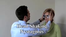 |
Head-Shaking Nystagmus | Head-shaking nystagmus: With a peripheral lesion, similar to vibration, transiently accentuates vestibular asymmetry when baseline VOR function is asymmetric, central patterns are well described and have localizing value (e.g., causing vertical nystagmus after horizontal head-shaking, horizontal nys... | Image/MovingImage |
| 104 |
 |
Head-Shaking Nystagmus - A 'Central' Pattern | 𝗢𝗿𝗶𝗴𝗶𝗻𝗮𝗹 𝗗𝗲𝘀𝗰𝗿𝗶𝗽𝘁𝗶𝗼𝗻: Evaluating for nystagmus provoked by head-shaking, so-called head-shaking nystagmus (HSN), should be performed in all patients with complaints of dizziness or vertigo, regardless of the chronicity. The maneuver is perform... | Image/MovingImage |
| 105 |
 |
Head-Shaking-Induced Nystagmus Following Ramsay Hunt Vestibulopathy | 𝗢𝗿𝗶𝗴𝗶𝗻𝗮𝗹 𝗗𝗲𝘀𝗰𝗿𝗶𝗽𝘁𝗶𝗼𝗻: This is a 50-year-old man who experienced the abrupt onset of imbalance, dizziness and left-sided hearing loss 4 months prior to this examination. He was found to have herpetic vesicles in the left external auditory canal... | Image/MovingImage |
| 106 |
 |
Hemifacial Spasm | This is a 45-year-old man with intermittent left facial twitching and eyelid closure for the last 6 months. With observation, spontaneous left facial spasms were seen involving the orbicularis oculi and oris muscles. With voluntary contraction of left facial muscles, with smiling for instance, there... | Image/MovingImage |
| 107 |
 |
HINTS Exam and Saccadic Dysmetria in Lateral Medullary Stroke | 𝗢𝗿𝗶𝗴𝗶𝗻𝗮𝗹 𝗗𝗲𝘀𝗰𝗿𝗶𝗽𝘁𝗶𝗼𝗻: This is a 50-year-old who experienced the abrupt onset of prolonged vertigo following chiropractic therapy 2 months prior. Initial work-up included an MRI and MR angiogram - MR-diffusion weighted imaging showed an acute l... | Image/MovingImage |
| 108 |
 |
Horizontal Canal - BPPV: BBQ Roll to Treat the Right Side | 𝗢𝗿𝗶𝗴𝗶𝗻𝗮𝗹 𝗗𝗲𝘀𝗰𝗿𝗶𝗽𝘁𝗶𝗼𝗻: To treat right horizontal canal (HC)-BPPV (each position maintained for at least 30 seconds or until nystagmus and/or vertigo cease): • First the patient is placed in the long-sitting position • Then in a supine posit... | Image/MovingImage |
| 109 |
 |
Horizontal Canal - BPPV: Gufoni for Right Apogeotropic | 𝗢𝗿𝗶𝗴𝗶𝗻𝗮𝗹 𝗗𝗲𝘀𝗰𝗿𝗶𝗽𝘁𝗶𝗼𝗻: To treat the right apogeotropic (beating towards the sky with right ear down and with left ear down - e.g., left beating nystagmus with right supine roll test or with right ear down; right beating nystagmus with left supi... | Image/MovingImage |
| 110 |
 |
Horizontal Gaze Palsy, Facial Nerve Palsy, and Nystagmus Due to Dorsal Pontine Ischemia | 𝗢𝗿𝗶𝗴𝗶𝗻𝗮𝗹 𝗗𝗲𝘀𝗰𝗿𝗶𝗽𝘁𝗶𝗼𝗻: Presented here are two patients with horizontal gaze and facial palsies due to stroke. The first patient is a 60-year-old man who presented with double vision and hemiparesis due to a right dorsal pontine ischemic stroke.... | Image/MovingImage |
| 111 |
 |
Horner's Syndrome with Anhidrosis | 𝗢𝗿𝗶𝗴𝗶𝗻𝗮𝗹 𝗗𝗲𝘀𝗰𝗿𝗶𝗽𝘁𝗶𝗼𝗻: This is a patient with the onset of ptosis OD years prior, with clear evidence of a Horner's syndrome. Imaging of the oculosympathetic tract was unrevealing. The patient also mentioned that with exercise, the left side of... | Image/MovingImage |
| 112 |
 |
How to Measure Ocular Alignment Virtually | Ocular alignment: the alternate cover test can be performed by instructing the patient to hold their head steady, fix their eyes on the camera (or a more distant target - the closer the fixation target, the more of an exodeviation the examiner will see), and use their cell phone (or a spoon) to occl... | Image/MovingImage |
| 113 |
 |
Hyperventilation | Hyperventilation: instruct the patient to breathe rapidly in and out of their mouth for 40-60 seconds. Alkalosis and changes in ionized calcium may improve conduction through an affected segment of 8th cranial nerve due to vestibular schwannoma (https://collections.lib.utah.edu/details?id=1213447) o... | Image/MovingImage |
| 114 |
 |
Hyperventilation-Induced Downbeat Nystagmus in a Cerebellar Disorder | 𝗢𝗿𝗶𝗴𝗶𝗻𝗮𝗹 𝗗𝗲𝘀𝗰𝗿𝗶𝗽𝘁𝗶𝗼𝗻: This is a 45-year-old woman with a chronic progressive cerebellopathy of unclear etiology (worsening over at least 10 years) characterized by gait and limb ataxia, gaze-evoked nystagmus, saccadic pursuit and vestibulo-ocu... | Image/MovingImage |
| 115 |
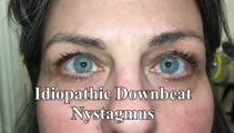 |
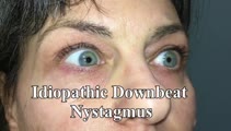
Idiopathic Downbeat Nystagmus Exacerbated with Positional Maneuvers | 𝗢𝗿𝗶𝗴𝗶𝗻𝗮𝗹 𝗗𝗲𝘀𝗰𝗿𝗶𝗽𝘁𝗶𝗼𝗻: This is a 45-yo-woman with vertical oscillopsia for 6+ months, found to have downbeat nystagmus on examination. She mainly complained of dizziness and oscillopsia when laying down. She was found to have a significant exac... | Image/MovingImage |
| 116 |
 |
Idiopathic Downbeat Nystagmus Exacerbated with Positional Maneuvers - Part 2: Patient is Now on 4-Aminopyridine | This is a 45-yo-woman presented in "Idiopathic downbeat nystagmus exacerbated with positional maneuvers". This video was taken after the patient had been on 4-aminopyridine for 3 months. There was marked improvement in subjective oscillopsia and objective downbeat nystagmus. The strong positional co... | Image/MovingImage |
| 117 |
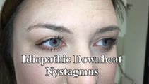 |
Idiopathic Downbeat Nystagmus, Decreasing with Convergence | This is a 25-yo-woman who experienced vertically oscillopsia for 1 year, and was found to have downbeat nystagmus. Interestingly, there were no other cerebellar ocular motor signs - e.g., normal saccades, smooth pursuit, VOR suppression, and no gaze-evoked nystagmus, although her (pure) downbeat was... | Image/MovingImage |
| 118 |
 |
Impaired Smooth Pursuit and Other Characteristic Ocular Motor Findings in Middle Cerebellar Peduncle Stroke | 𝗢𝗿𝗶𝗴𝗶𝗻𝗮𝗹 𝗗𝗲𝘀𝗰𝗿𝗶𝗽𝘁𝗶𝗼𝗻: This is a 50-year-old woman who underwent resection of a left-sided acoustic neuroma, and post-operatively, she had vertigo, binocular diplopia, left hemi-ataxia and severe gait ataxia. MR diffusion weighted imaging demon... | Image/MovingImage |
| 119 |
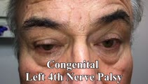 |
Inferior Oblique Overaction in a Congenital 4th Nerve Palsy | 60-yo-man complaining of intermittent oblique diplopia. There was a left hypertropia that worsened in down gaze, right gaze and in left head tilt. There was a large vertical fusional amplitude in addition to a longstanding rightward head tilt, and on examination there was left inferior oblique overa... | Image/MovingImage |
| 120 |
 |
The Influence of Convergence on Downbeat Nystagmus | This is a patient presenting with progressive imbalance and oscillopsia over the course of approximately 1 year. On examination, he had cerebellar ataxia in addition to spontaneous downbeat nystagmus (DBN). His downbeat nystagmus increased in lateral and downgaze, which are characteristic features,... | Image/MovingImage |
| 121 |
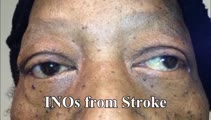 |
INOs in Stroke | 𝗢𝗿𝗶𝗴𝗶𝗻𝗮𝗹 𝗗𝗲𝘀𝗰𝗿𝗶𝗽𝘁𝗶𝗼𝗻: This video shows 3 patients with vascular risk factors who suffered strokes of the MLF resulting in unilateral INO in each case. In the second case, INO was diagnosed status post cardiac catherization and MRI was found to... | Image/MovingImage |
| 122 |
 |
Internuclear Ophthalmoplegia (INO) in Multiple Sclerosis | 𝗢𝗿𝗶𝗴𝗶𝗻𝗮𝗹 𝗗𝗲𝘀𝗰𝗿𝗶𝗽𝘁𝗶𝗼𝗻: This video includes 3 patients each with a known history of MS found to have unilateral or bilateral INOs on their exam. In the first 2 patients, the INOs are relatively subtle with normal adduction. However, with rapid h... | Image/MovingImage |
| 123 |
 |
Ipsitorsional Quick Phases with Head Tilt in a Normal Subject | This is a demonstration of ocular counterroll, which can be seen when the head is tilted to the right or to the left. For example, when the head is tilted to the right, the top poles of both eyes should rotate toward the left ear to keep the top poles oriented with earth vertical. This is part of ... | Image/MovingImage |
| 124 |
 |
Isolated Central 4th Nerve Palsy | 𝗢𝗿𝗶𝗴𝗶𝗻𝗮𝗹 𝗗𝗲𝘀𝗰𝗿𝗶𝗽𝘁𝗶𝗼𝗻: This is a 40-year-old man with a right hypertropia that worsened in left and down gaze in addition to right head tilt, and improved in left head tilt. There was subjective excyclotorsion OD with double Maddox rod testing.... | Image/MovingImage |
| 125 |
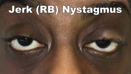 |
Jerk Nystagmus | 𝗢𝗿𝗶𝗴𝗶𝗻𝗮𝗹 𝗗𝗲𝘀𝗰𝗿𝗶𝗽𝘁𝗶𝗼𝗻: This is an example of jerk nystagmus due to a central vestibular lesion. The slow phase is the pathologic phase (to the left) which initiates the movement, and is followed by a fast position reset mechanism (to the right)... | Image/MovingImage |