AAO-NANOS Neuro-Ophthalmology Clinical Collection: Derived from the AAO-NANOS Clinical Neuro-Ophthalmology collection produced on CD. The images are of selected cases from the NANOS teaching slide exchange, and the CD was produced under the direction of Larry Frohman, MD and Andrew Lee, MD.
The American Academy of Ophthalmology (AAO); The North American Neuro-Ophthalmology Association (NANOS).
NOVEL: https://novel.utah.edu/
TO
| Title | Creator | Description | ||
|---|---|---|---|---|
| 101 |
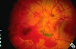 |
Isolated Optic Neuritis/Neuropathy | Rosa A. Tang, MD | Papilledema may produce visual loss due to chronic atrophic papilledema, secondary macular hemorrhage, exudate or edema, secondary ischemic optic neuropathy, or secondary subretinal neovascular membrane formation. |
| 102 |
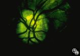 |
Optic Disc Drusen With Autofluorescence | Thomas R. Wolf, MD | This photograph of optic disc drusen demonstrates autoflourescence with flourescein barrier filters in place. Imaging: flourescein barrier filters. |
| 103 |
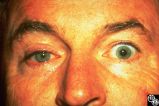 |
Migraine Syndrome | Mitchell J. Wolin, MD | The image shows a patient with cluster headache and eye displaying Horner's syndrome. |
| 104 |
 |
Isolated Congenital Optic Disc Anomalies | Thomas R. Wolf, MD | Benign tumors of blood vessels (hemangiomas) may occur on the optic nerve and may mimic optic disc edema. Disease/Diagnosis: Optic Nerve Hemangioma. |
| 105 |
 |
Motility Disturbances | Don Bienfang, MD | This patient displays a posttraumatic left fourth nerve palsy sustained after having struck her head on the dashboard. |
| 106 |
 |
Isolated Optic Neuritis/Neuropathy | Rosa A. Tang, MD | Papilledema in pseudotumor cerebri may result in adjacent choroidal or retinal folds. |
| 107 |
 |
Optic Tract Syndrome Due to Carotid Artery Dolichoectasia | Larry P. Frohman, MD | This 43-year-old man was referred for evaluation of 6 months of visual loss OU. In retrospect, he had noticed increasing difficulty with his tennis game dating back over 3 years, as balls would pass him unexpectedly when hit to his backhand (left) side. The patient did not think this was progressive... |
| 108 |
 |
Optic Tract Syndrome Due to Carotid Artery Dolichoectasia | Larry P. Frohman, MD | This 43-year-old man was referred for evaluation of 6 months of visual loss OU. In retrospect, he had noticed increasing difficulty with his tennis game dating back over 3 years, as balls would pass him unexpectedly when hit to his backhand (left) side. The patient did not think this was progressive... |
| 109 |
 |
Isolated Congenital Optic Disc Anomalies | Rosa A. Tang, MD | This patient has optic disc drusen and evidence of a superimposed optic neuropathy, including loss of visual field, an ipsilateral afferent pupillary defect, and optic atrophy. Although optic disc drusen typically causes visual field loss without visual acuity loss superimposed, ischemic optic neuro... |
| 110 |
 |
Orbital Tumors | Mitchell J. Wolin, MD | Cavernous hemangiomas of the orbit usually result in painless orbital signs such as proptosis or visual loss. Orbital imaging of the lesion, which usually is a well-defined orbital mass, is demonstrated in this study. The lesion is benign and usually occurs in young to middle-aged adults. Surgical e... |
| 111 |
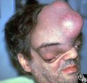 |
Neuro-Ophthalmic Manifestations of Brain Tumors | Steven Galetta, MD | The patient is a 45-year-old recluse found to harbor this frontal lobe mass. Remarkably, this patient had only mild bilateral optic neuropathies with visual acuities in the 20/25 range. This right disc was mildly swollen and the left mildly pale. He could not fit into the fundus camera for disc phot... |
| 112 |
 |
Motility Disturbances | Rosa A. Tang, MD | Skew deviation is a vertical deviation that is not localized to any one muscle or muscle group. The deviation may be comitant or not, and intermittent or constant. Skew deviation is often defined by the company it keeps, that is, skew usually occurs in association with other brain-stem signs, and is... |
| 113 |
 |
Motility Disturbances | Rosa A. Tang, MD | Skew deviation is a vertical deviation that is not localized to any one muscle or muscle group. The deviation may be comitant or not, and intermittent or constant. Skew deviation is often defined by the company it keeps, that is, skew usually occurs in association with other brain-stem signs, and is... |
| 114 |
 |
Pupillary Syndrome | Jeffrey G. Odel, MD | Intermittent dilation of the pupils may occur as a benign phenomenon in healthy young adults. In the absence of other third nerve signs, (eg, ptosis, diplopia, ophthalmoplegia), an isolated transient dilation of the pupil in an otherwise healthy adult is unlikely to represent a third nerve palsy. Tr... |
| 115 |
 |
Pupillary Syndrome | Jeffrey G. Odel, MD | Intermittent dilation of the pupils may occur as a benign phenomenon in healthy young adults. In the absence of other third nerve signs, (eg, ptosis, diplopia, ophthalmoplegia), an isolated transient dilation of the pupil in an otherwise healthy adult is unlikely to represent a third nerve palsy. Tr... |
| 116 |
 |
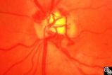
Acquired Disc Changes | Rosa A. Tang, MD | Although optociliary shunt vessels are venous collaterals that form in response to chronic venous obstruction, they may occur in patients with chronic open-angle glaucoma. |
| 117 |
 |
Acquired Disc Changes | Rosa A. Tang, MD | Although optociliary shunt vessels are venous collaterals that typically form in response to chronic venous obstruction, they may occur on a congenital basis as seen here. |
| 118 |
 |
Isolated Optic Neuritis/Neuropathy | Rosa A. Tang, MD | Papilledema is a term reserved for optic disc edema related to increased intracranial pressure. Fluid within the optic nerve sheath or elevation of the intraocular optic nerve head may be visible on magnetic resonance imaging studies of the head and orbit. |
| 119 |
 |
Neuro-Ophthalmic Imaging-MRI | Rosa A. Tang, MD | Aneurisms may result in neuro-ophthalmologic sign and symptoms by direct compression of the afferent or efferent systems or by the secondary effects of hemorrhage. Basilar aneurisms may result in ocular motor deficits such as a unilateral or bilateral third nerve palsy. |
| 120 |
 |
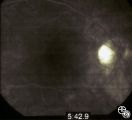
Acquired Disc Changes | Rosa A. Tang, MD | Optociliary shunt vessels are venous collaterals that form in response to chronic venous obstruction. They may occur in patients following central retinal vein occlusion. |
| 121 |
 |
Isolated Optic Neuritis/Neuropathy | Larry P. Frohman, MD | The patient is a 62-year-old female who presented in August 1996 with visual loss OD that she first noted as loss of her superior field in May 1996. She felt that it had been static since, and perhaps was even a little better in the week before she was seen. There was no pain, even with ocular rotat... |
| 122 |
 |
Isolated Optic Neuritis/Neuropathy | Larry P. Frohman, MD | The patient is a 62-year-old female who presented in August 1996 with visual loss OD that she first noted as loss of her superior field in May 1996. She felt that it had been static since, and perhaps was even a little better in the week before she was seen. There was no pain, even with ocular rotat... |
| 123 |
 |
Isolated Optic Neuritis/Neuropathy | Larry P. Frohman, MD | The patient is a 62-year-old female who presented in August 1996 with visual loss OD that she first noted as loss of her superior field in May 1996. She felt that it had been static since, and perhaps was even a little better in the week before she was seen. There was no pain, even with ocular rotat... |
| 124 |
 |
Isolated Optic Neuritis/Neuropathy | Larry P. Frohman, MD | The patient is a 62-year-old female who presented in August 1996 with visual loss OD that she first noted as loss of her superior field in May 1996. She felt that it had been static since, and perhaps was even a little better in the week before she was seen. There was no pain, even with ocular rotat... |
| 125 |
 |
Isolated Optic Neuritis/Neuropathy | Larry P. Frohman, MD | The patient is a 62-year-old female who presented in August 1996 with visual loss OD that she first noted as loss of her superior field in May 1996. She felt that it had been static since, and perhaps was even a little better in the week before she was seen. There was no pain, even with ocular rotat... |