The Health Education Assets Library (HEAL) is a collection of over 22,000 freely available digital materials for health sciences education. The collection is now housed at the University of Utah J. Willard Marriott Digital Library.
TO
Filters: Collection: ehsl_heal
| Title | Description | Subject | Collection | ||
|---|---|---|---|---|---|
| 76 |
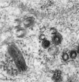 |
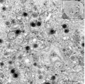
Mast cell (connective tissue, human) | Electron microscopy. Mast cells (mastocytes) are frequently found perivascularly or perineurally within the connective tissue. A detail of this mast cell shows granules varying in shape and size. These membrane-bound vesicles (so-called compound granules) show a metachromatic reaction in light micro... | Poja Histology Collection - Blood & Bone Marrow Subset | |
| 77 |
 |
Mast cell (lung, dog) | Electron microscopy. This mucosal mast cell of the lung is localized in the vicinity of a blood vessel. Notice the smooth muscle cells (1) of a small arteriole and collagen fibers (2). Most obvious is the presence of granules varying in shape and size. These membrane-bound vesicles (so-called compou... | Poja Histology Collection - Blood & Bone Marrow Subset | |
| 78 |
 |
Mast cell (lung, golden hamster) | Electron microscopy. Mucosa mast cells (mastocytes) are frequently found perivascularly or perineurally. The mast cell shows long thin microvilli at the surface and the cytoplasm is stuffed with granules (1) varying in shape and size. At (2) a Golgi area and at (3) small mitochondria and some electr... | Poja Histology Collection - Blood & Bone Marrow Subset | |
| 79 |
 |
Mast cell (lung, human) | Electron microscopy. Mast cells (mastocytes) are oval (12 m) or spindle-shaped and show few thin microvilli at the surface. They are frequently found perivascularly or perineurally within the connective tissue (1). The cytoplasm is provided with a moderate amount of organelles. Most obvious is the p... | Poja Histology Collection - Blood & Bone Marrow Subset | |
| 80 |
 |
Mast cell (lung, human) | Electron microscopy. Mast cells (mastocytes) are frequently found perivascularly or perineurally. A part of this mast cell shows thin microvilli at the surface and the cytoplasm is provided with a moderate amount of organelles. At (*) the Golgi area (1) is seen in close association with granules tha... | Poja Histology Collection - Blood & Bone Marrow Subset | |
| 81 |
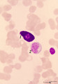 |
Mast cell in bone marrow smear (human) | Stain: May-Grnwald-Giemsa (MGG). The mast cell (1) is characterized by the presence of coarse purple-black granules and a nucleus with oval, central nucleus. The granules contain pharmacologically active mediators such as heparin, histamine, neutrophil- and eosinophil-chemotactic factors, vasoactive... | Poja Histology Collection - Blood & Bone Marrow Subset | |
| 82 |
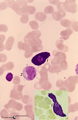 |
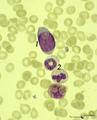
Mast cells in peripheral blood smear (human) | Stain: May-Grnwald-Giemsa (MGG). Mast cells (1) from two different smears. (2) Monocyte. Mast cells or tissue basophils are normally present in small numbers in bone marrow smears. They must not be confused with developing basophils. Mast cells are large cells (20-30 m in diameter) with an irregular... | Poja Histology Collection - Blood & Bone Marrow Subset | |
| 83 |
 |
Maturation stages of plasmablasts (spleen, liver rat) | Electron microscopy. The youngest (plasmacytoid) lymphocyte in (A) shows a slight indented nucleus with a distinct nucleolus. Apart from a few mitochondria the cytoplasm contains large amount of free ribosomes and polysomes, few profiles of long rough endoplasmic reticulum (1, RER) and Golgi areas (... | Poja Histology Collection - Blood & Bone Marrow Subset | |
| 84 |
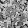 |
Mature erythrocytes (spleen, gerbil) | Scanning electron microscopy. Normal mature erythrocytes (1) are mostly biconcave and discoid. They might change their forms due to mechanical pressures. (2) a mature lymphocyte recognizable by many short stubby microvilli at the surface. (3) granulocytes are larger with fewer but longer microvilli.... | Poja Histology Collection - Blood & Bone Marrow Subset | |
| 85 |
 |
Megakaryoblast in bone marrow smear (human) | Stain: May-Grnwald-Giemsa (MGG). The megakaryoblast (1) is not yet producing platelets and has a relative small amount of basophilic cytoplasm. The nucleus is large with fine disperse chromatin and nucleoli. Most other nucleated cells are of the myeloid cell line. | Poja Histology Collection - Blood & Bone Marrow Subset | |
| 86 |
 |
Megakaryocyte | Scheme electron microscopy. The megakaryocyte is derived from a pro-megakaryocyte which originates from splenic stem cells (CFU-S, colony forming units-spleen). The megakaryocyte is a giant polyploid cell (35-160 m) and contains a large multilobate nucleus (1). The perinuclear area shows Golgi areas... | Poja Histology Collection - Blood & Bone Marrow Subset | |
| 87 |
 |
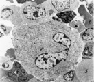
Megakaryocyte (bone marrow, mouse) | Electron microscopy. A detail of the cytoplasm of a megakaryocyte (see inset) demonstrates distinctly a part of the intermediate zone subdivided by an interconnected tubular system (so-called demarcation membrane system or open canalicular system: OCS) that is in continuity with the cell surface. Th... | Poja Histology Collection - Blood & Bone Marrow Subset | |
| 88 |
 |
Megakaryocyte (bone marrow, rabbit) | Electron microscopy. A survey of a megakaryocyte demonstrates very well the differences in diameter between this giant polyploid cell (1) and the other young white blood cells (2-5). In the intermediate zone the (dark) granules (of the future platelets) are close associated with the electron-light i... | Poja Histology Collection - Blood & Bone Marrow Subset | |
| 89 |
 |
Megakaryocyte (peripheral blood, human) | Electron microscopy. A small part of a megakaryocyte (see also inset) shows two nuclear segments (1) and two areas of cytoplasm. Seen at this magnification and close to the perinuclear area, the granular population can be divided into homogeneous electron-dense (2) and electron-grey (3) granules. Th... | Poja Histology Collection - Blood & Bone Marrow Subset | |
| 90 |
 |
Megakaryocyte in bone marrow smear (human) | Stain: May-Grnwald-Giemsa (MGG), (black and white print). The giant polyploid cell (35-160 μm) contains a large multilobate nucleus. Note the ring-like pattern of the nuclei (1). The light-stained perinuclear zone (2) contains Golgi areas, within the encompassing greyish (intermediate) zone (3) gra... | Poja Histology Collection - Blood & Bone Marrow Subset | |
| 91 |
 |
Mitosis in bone marrow smear (human) | Stain: May-Grnwald-Giemsa (MGG). Increased mitotic activity can be observed in megaloblastic anemia. (1) Mitosis figure. (2) Basophilic erythroblasts. | Poja Histology Collection - Blood & Bone Marrow Subset | |
| 92 |
 |
Monoblast | Scheme electron microscopy. The precursor of this cell is the promonoblast derived from the common stem cell CFU-GM (colony forming unit-granulocyte/monocyte). After proliferation of the promonoblasts they transform into monoblasts (up to 10 μm). The nucleus of the monoblast shows an indentation as... | Poja Histology Collection - Blood & Bone Marrow Subset | |
| 93 |
 |
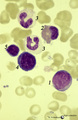
Monoblast in bone marrow smear (human) | Stain: May-Grnwald-Giemsa (MGG). The monoblast (1) has a large nucleus with an irregular border, nucleoli and fine disperse chromatin, and a basophilic cytoplasm with no or a limited number of granules. (2) Two neutrophilic band forms. | Poja Histology Collection - Blood & Bone Marrow Subset | |
| 94 |
 |
Monoblast, erythroblasts and granulopoietic cells in bone marrow smear (human) | Stain: May-Grnwald-Giemsa (MGG). The monoblast (1) has a large nucleus with an irregular border, nucleoli and fine disperse chromatin, and a basophilic cytoplasm (greyish blue) with no or hardly any granules. (2) Eosinophilic metamyelocyte with large, brown granules. (3) Juvenile unsegmented (band f... | Poja Histology Collection - Blood & Bone Marrow Subset | |
| 95 |
 |
Monoblast, myeloid and erythropoietic cells in bone marrow smear (human) | Stain: May-Grnwald-Giemsa (MGG). (1) late promyelocyte or early myelocyte with nucleoli in the nucleus and ample azurophilic granules. (2) monoblast with indented nucleus and nucleoli (3) polychromatic erythroblast that will turn into an orthochromatic erythroblast (4), with progressing chromatin co... | Poja Histology Collection - Blood & Bone Marrow Subset | |
| 96 |
 |
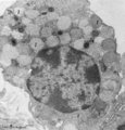
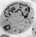
Monocyte | Scheme electron microscopy. The diameter of monocytes ranges from 12-20 m. Characteristic is the large (kidney-shaped) nucleus with one or more nucleoli (1) and several indentations. The cytoplasm contains: (2) Golgi areas, (3) electron-dense lysosomal granules or primary or azurophilic granules wit... | Poja Histology Collection - Blood & Bone Marrow Subset | |
| 97 |
 |
Monocyte (peripheral blood, human) | Electron microscopy. In this picture the large nucleus (1) of this cell (diameter of 12-20 μm) is twice sectioned. Golgi areas (2) and centriole, profiles of rough endoplasmic reticulum (3) and many free ribosomes are present. There are many mitochondria (4) as well as scattered homogeneous electro... | Poja Histology Collection - Blood & Bone Marrow Subset | |
| 98 |
 |
Monocyte (peripheral blood, human) | Electron microscopy. In this picture the large nucleus of this cell (diameter of 12-20 μm) is twice sectioned (1). Golgi areas (2), a few profiles of rough endoplasmic reticulum (3) and many free ribosomes are present. There are many mitochondria (4) as well as small-sized light vesicles (5). The c... | Poja Histology Collection - Blood & Bone Marrow Subset | |
| 99 |
 |
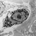
Monocyte (peripheral blood, human) | Electron microscopy. A large cell with an indented nucleus (1), few Golgi areas (2) and numerous organelles. The cytoplasm contains scattered homogeneous electron-dense lysosomal granules (3) (azurophilic granules with acid phosphatase, arylsulfatase) in variable amounts and few vacuoles. Small pseu... | Poja Histology Collection - Blood & Bone Marrow Subset | |
| 100 |
 |
Monocyte in peripheral blood smear (human) | Stain: May-Grnwald-Giemsa (MGG). The monocyte (12-20 μm) contains a large light-stained nucleus with a characteristic indentation with a distinct nucleolus in a light blue-stained apparently transparent cytoplasm. Small primary or azurophilic granules are hardly visible. | Poja Histology Collection - Blood & Bone Marrow Subset |