The Health Education Assets Library (HEAL) is a collection of over 22,000 freely available digital materials for health sciences education. The collection is now housed at the University of Utah J. Willard Marriott Digital Library.
Filters: Collection: ehsl_heal
| Title | Description | Subject | Collection | ||
|---|---|---|---|---|---|
| 76 |
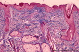 |
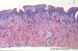
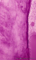
Lip (human), skin surface. | Stain: Azan. Keratinized squamous epithelium with hair follicles; submucosa with sebaceous glands, and extensions of skeletal muscle cells (orbicularis oris). | oral cavity | Poja Histology Collection - Oral Cavity Subset |
| 77 |
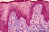 |
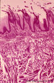
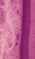
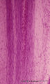
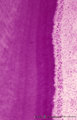
Lip (human), transitional zone (red zone or vermilion border) | Stain: Azan. Slightly cornified epithelium with high irregular dermal papillae and many capillaries. Note the epithelium is thicker, but less cornified than the epidermis. The red color of the lips is due to the rich vascularity of the lamina propria and the lucidity of the epithelium. | oral cavity; lining mucosa; red zone; vermilion border | Poja Histology Collection - Oral Cavity Subset |
| 78 |
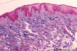 |
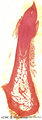
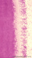
Lip (human), transitional zone (red zone or vermilion border) | Stain: Azan. Slightly cornified epithelium with high irregular dermal papillae and many capillaries. Fat cells and skeletal muscle cells are located in the submucosa. | oral cavity; lining mucosa; red zone; vermilion border | Poja Histology Collection - Oral Cavity Subset |
| 79 |
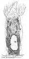 |
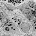
Odontoblast in tooth development - mammalian embryo | Scheme electronmicroscopy. A columnar and irregular shaped cell (mesenchymal origin) with a taperwise odontoblastic process within the predentin. The bottom side is adjacent to the future pulp cells. The cell body contains many organelles and especially a well-developed Golgi area with prosecretoy g... | oral cavity | Poja Histology Collection - Oral Cavity Subset |
| 80 |
 |
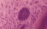
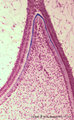
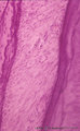
Papillae circumvallatae of the tongue (dorsal side, human) | Stain: Azan. Three broad papillae with taste buds facing the grooves in which the serous von Ebner glands drain. Striated skeletal muscles (musculus verticalis linguae and musculus longitudinalis superior) and lightly stained mucous glands (posterior lingual glands). | oral cavity; von Ebner; lingual muscles; lingual glands | Poja Histology Collection - Oral Cavity Subset |
| 81 |
 |
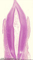
Papillae circumvallatae of the tongue (dorsal side, human) | Stain: Azan. A broad papilla with taste buds (lightly stained spots) facing the grooves in which the serous glands (von Ebner) drain. | oral cavity; von Ebner; circumvallate papillae | Poja Histology Collection - Oral Cavity Subset |
| 82 |
 |
Papillae filliformes of the tongue (dorsal side, human) | Stain: Heidenhain light bordeaux. Threadlike keratinized extensions of the stratified epithelium. Primary connective tissue papillae with 2 to 3 secondary papillae. The skeletal muscle fibers are arranged in three directions. | oral cavity; filiform papillae | Poja Histology Collection - Oral Cavity Subset |
| 83 |
 |
Parotid gland (human) | Stain: Mallory trichrome. Survey: at the left bottom a large interlobular duct (in lumen remnants of secretion products) within a septum of dense connective tissue. At the top thinner (blue) septum, a thick (red-bluish) septum at the left. In the center three (intralobular) striated ducts between th... | oral cavity; serous gland | Poja Histology Collection - Oral Cavity Subset |
| 84 |
 |
Parotid gland (human) | Stain: Azan. The parotid gland: in most species the gland is composed entirely of serous acini. At the right a small (intralobular) striated duct; centrally one large interlobular duct with blood vessels. Scattered a few (white) fat cells. | oral cavity; serous gland | Poja Histology Collection - Oral Cavity Subset |
| 85 |
 |
Parotid gland (rat) | Electronmicroscopy. Part of a serous acinus with characteristic secretion granules supranuclearly. Note different densities of the granules without any signs of fusion. | oral cavity; serous gland | Poja Histology Collection - Oral Cavity Subset |
| 86 |
 |
Periodontal ligament with epithelial rest of Malassez - longitudinal section of root of tooth, higher magnification; human, adult | Stain: Hematoxylin and eosin. Centrally within the connective tissue of the periodontal ligament a distinct darker stained epithelial islet with nuclei is present (epithelial rest of Malassez as a remnant of Hertwig's epithelial root sheath; in adults it might produce dental cyst). At the right side... | oral cavity; cementoblasts; epithelial rest of Malassez; cementoid | Poja Histology Collection - Oral Cavity Subset |
| 87 |
 |
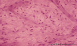
Periodontal ligament with epithelial rests of Malassez - longitudinal section of root of tooth; human, adult | Stain: Hematoxylin and eosin. From left to right: connective tissue of periodontal ligament with epithelial rests of Malassez as the persistent remnants of the epithelium of Hertwig's epithelial root sheath. The three islets close to the cemental zone are slightly darker stained (note cluster of nuc... | oral cavity; cementoblasts; Sharpey's fibers; acellular cementum | Poja Histology Collection - Oral Cavity Subset |
| 88 |
 |
Permanent tooth - canine, human, adult; low magnification of labiolingual section | Stain: Hematoxylin and scarlet red. From top to bottom: Crown region with dentin but without enamel (decalcified specimens); Neck region at the attachment of the gingiva to dentin (left and right); Cementum is visible as a dark small rim from the neck region to the bottom of the tooth; Periodontal l... | oral cavity; alveolar process | Poja Histology Collection - Oral Cavity Subset |
| 89 |
 |
Predentin formation at the cuspal tip in tooth development - bell stage, human, embryo | Stain: Azan. From top to bottom: Stellate reticulum consisting of a network of ectoderm-derived cells; Cell layers of the stratum intermedium; Columnar (presecretory) ameloblasts with their upper side (nuclear area) in close contact with the stratum intermedium, and at the distal side (secretion are... | oral cavity; predentin | Poja Histology Collection - Oral Cavity Subset |
| 90 |
 |
Premolar permanent tooth (human, adult; low magnification of labiolingual section) | Stain: Hematoxylin and eosin. From top to bottom: Crown region with enamel-dentin boundary (there is no enamel present in decalcified specimens); dentin (blue-pink) enwraps the whole pulp chamber (light) and here the lines of dentinal tubules appear S-shaped. Neck region at the attachment of the gin... | oral cavity; pulp canal | Poja Histology Collection - Oral Cavity Subset |
| 91 |
 |
Presecretory ameloblast in tooth development - mammalian embryo | Scheme electronmicroscopy. The ectodermal-derived cell appears as tall columnar with fingerlike extensions (dependent on the development stages) at their distal side (secreting area = 'functional base'). These extensions are formed as the cell withdraws during the production of initial enamel. The s... | oral cavity | Poja Histology Collection - Oral Cavity Subset |
| 92 |
 |
Presecretory ameloblasts in tooth development - bell stage, gerbil, postnatal | Electronmicroscopy. Well-arranged epithelial formation of presecretory ameloblasts (active nuclei) with their distal secretion sides towards the thin grey basal lamina. Predentin at the bottom close to the basal lamina and comprises collagen fibers, odontoblastic extensions and dispersed calcified m... | oral cavity; predentin; matrix vesicles | Poja Histology Collection - Oral Cavity Subset |
| 93 |
 |
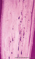
Pulp canal of tooth - longitudinal section; human, adult | Stain: Hematoxylin and eosin. Centrally three thick nerve bundles. In between blood vessel filled with erythrocytes. At the right side longitudinal course of slender thin-walled blood vessels (arterioles) which pass into thin-walled capillaries with a wide lumen. | oral cavity; pulp canal | Poja Histology Collection - Oral Cavity Subset |
| 94 |
 |
Pulp chamber of tooth - human, adult | Stain: Hematoxylin and eosin. Pulpal connective tissue is a special type of tissue (stromal tissue) with so-called stellate-shaped pulpal fibroblastic cells that produce growth factors, extracellular matrix including thin collageneous proteins. Blood vessels are small and thin-walled; macrophages an... | oral cavity; pulp chamber; pulp fibroblast | Poja Histology Collection - Oral Cavity Subset |
| 95 |
 |
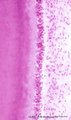
Pulp stones in root of tooth - longitudinal section, human, adult | Stain: Hematoxylin and eosin. Left and right side dark stained dentin with a small light rim of predentin. Centrally pulp stones represent as free lying irregular calcifications. They are found diffusely distributed as spots or as thin strands following blood vessels and bundles of collagen fibers i... | oral cavity; pulp stone; diffuse calcifications | Poja Histology Collection - Oral Cavity Subset |
| 96 |
 |
Pulpo-dentinal complex - segment of a tooth; human, adult | Stain: Hematoxylin and eosin. At the pulp-dentin border the peripheral pulp appears to be a specialized odontogenic region composed of odontoblasts, a cell-free zone and a cell-rich zone. From left to right: dentin (note striation due to dentinal tubules); (uncalcified) light stained predentin. At t... | oral cavity; Tomes' Fibers; dentinal tubules; predentin | Poja Histology Collection - Oral Cavity Subset |
| 97 |
 |
Pulpo-dentinal complex in longitudinal section of tooth - damaged dentin, human, adult | Stain: Hematoxylin and eosin. If odontoblastic processes are damaged, (e.g., erosion, caries, etc.), the entire cell will degenerate, though it may be able to produce dentin. From left to right: - irregular course of dentinal tubules; - light stained (damaged) area contain no tubules; - pulp connect... | oral cavity | Poja Histology Collection - Oral Cavity Subset |
| 98 |
 |
Pulpo-dentinal complex in longitudinal section of tooth - secondary dentin, human, adult | Stain: Hematoxylin and eosin. From left to right: - dentinal tubules as fine striation in dentin; - vertical course of incremental lines (von Ebner); - a darker stained broad zone of secondary dentin; - pulp chamber with crowding of odontoblasts close to secondary, newly formed dentin; - connective ... | oral cavity; secondary dentin | Poja Histology Collection - Oral Cavity Subset |
| 99 |
 |
Pulpo-dentinal complex of the tooth (human, adult). | Stain: Hematoxylin and eosin. At the pulpo-dentinal border the peripheral pulp appears to be a specialized odontogenic region composed of odontoblasts, a cell-free zone and a cell-rich zone; from left to right: Dentin (note striation due to dentinal tubules). (Uncalcified) Light stained predentin. A... | oral cavity; Tomes' Fibers; dentinal tubules; predentin | Poja Histology Collection - Oral Cavity Subset |
| 100 |
 |
Root of tooth within osseous socket - cross-section; human, adult | Stain: Hematoxylin and eosin. From left to right: bundle bone of alveolus; oblique fibers of periodontal ligament with blood vessels (broad layer); flattened cementoblasts settled on a thin light rim of cementoid; darker stained acellular cementum, the so-called acellular extrinsic fiber cementum a... | oral cavity; acellular cementum | Poja Histology Collection - Oral Cavity Subset |