Collection of materials relating to neuro-ophthalmology as part of the Neuro-Ophthalmology Virtual Education Library.
NOVEL: https://novel.utah.edu/
TO
- NOVEL966
Filters: Collection: "ehsl_novel_novel"
| Title | Creator | Description | Subject | ||
|---|---|---|---|---|---|
| 76 |
 |
A Brief Introduction to Post-Ocular Surgery Strabismus | Srujay Pandiri, Medical Student | This is a short narrated Powerpoint that introduces concepts important to post-ocular surgery strabismus. It highlights the connection between common surgeries include cataract surgery, scleral buckle surgery, refractive surgery, among others. | Diplopia; Strabismus; Post-ocular Surgery Strabismus |
| 77 |
 |
My DEI Journey: Lessons Learned | Ore-ofe Adesina, MD | In this lecture, Dr. Adesina reviews his DEI journey in ophthalmology and the lessons of diversity, equity and inclusion he has learned along the way. He discusses why diversity, equity, and inclusion are all needed for a state of inclusive well-being, why representation matters for patient care and... | Ophthalmology; Diversity, Equity, Inclusion; Representation; Social Determinants of Health |
| 78 |
 |
Uveo-Meningeal Syndromes: Vogt-Koyanagi-Harada (VKH) Disease | Rachana Haliyur, MD, PhD; Emily Cole, MD, MPH; Therese Sassalos, MD; Sangeeta Khanna, MD | Ocular inflammatory symptoms with concurrent neuro-ophthalmologic manifestations can be diagnostically challenging. We provide a general overview of uveo-meningeal syndromes, which comprises a heterogeneous group of disorders that involve inflammation of the uveal tract, retina, and meninges. The pr... | VKH; Vogt-Koyanagi-Harada; Uveomeningeal Syndrome; Ocular Inflammation; Uveitis; CSF Pleocytosis; Serous Retinal Detachments |
| 79 |
 |
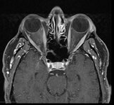
Metastatic Glioblastoma to Intracranial Optic Nerves, Optic Chiasm and Optic Tracts | Bashaer Aldhahwani, MD; Mariam S. Vilá-Delgado, MD | The patient with pathology confirmed glioblastoma after resectioning the superior frontal lobe tumor followed by 6 weeks of radiation therapy with concurrent temozolomide. The patient started bevacizumab to treat steroid-refractory vasogenic cerebral edema/radiation necrosis. 8 months after radiatio... | Metastatic Glioblastoma; Infiltrative Chiasmal Lesion |
| 80 |
 |
Unilateral Oculomotor Nerve Palsy Secondary to Internal Carotid Artery Aneurysm Without Pupil involvement: A Case Report | Danilo Andriatti Paulo; Richard J Blanch | Acquired oculomotor palsies (OMP) can result from numerous factors. The most common causes are presumed microvascular, trauma, compressive neoplasm, postneurosurgery and compression from aneurysm.1,2 ONP caused by internal carotid artery (ICA) aneurysm is a common clinical manifestation suggesting i... | Unilateral Oculomotor Nerve Palsy; Internal Carotid Artery Aneurysm; Pupil Involvement; Oculomotor Nerve Palsy; Secondary Oculomotor Nerve Palsy |
| 81 |
 |
Ethambutol Optic Neuropathy | Hailey Mair, BS; Padmaja Sudhakar, MD | This is a PowerPoint slide describing ethambutol induced optic neuropathy and it elaborates on mechanism and vulnearble population. | Ethambutol; Tuberculosis; Optic Neuropathy |
| 82 |
 |
Neuro-Ophthalmology of Multiple Sclerosis | Emily Sun, Medical Student; Amanda Henderson, MD | Multiple Sclerosis (MS) is the most common neurological disease in young people with an average age of onset between 15 and 35 years old. MS is an autoimmune inflammatory condition that causes demyelinating lesions in the CNS. The diagnosis is clinical, but MRI is typically used to support the diagn... | Multiple Sclerosis; Optic Neuritis; Uveitis; Internuclear Ophthalmoplegia; Nystagmus; Steroids; Demyelinating; Autoimmune |
| 83 |
|
Clival Menigioma Causing 6th Nerve Palsy | Bashaer Aldhahwani, MD; Mariam S. Vilá-Delgado, MD | 65 year- old Male patient presented with progressive binocular, horizontal diplopia with limitation of abduction in the right eye. He was diagnosed with Isolated compressive right 6th CN palsy due to right Clival Meningioma. | Clival Meningioma; Sixth Nerve Palsy |
| 84 |
 |
Diagnosis and Evaluation of Stroke: Mechanism - Vasculitis | Danny Alevy; Amanda D. Henderson, MD | In this video, we discuss the diagnosis and evaluation of several vasculitides linked to an increase in risk for cerebral ischemic stroke. We focus on ANCA-associated vasculitides, polyarteritis nodosa, Takayasu arteritis, lupus vasculitis, and Susac syndrome, while highlighting their key symptoms a... | Ischemic Stroke; Vasculitis; ANCA-associated Vasculitis; Polyarteritis Nodosa; Takayasu Arteritis; Systemic Lupus Erythematosus; Susac Syndrome |
| 85 |
 |
Illustrations of the Afferent Visual Pathway and Concepts Surrounding Trans-Synaptic Neuroaxonal Degeneration in the Visual Pathway in Multiple Sclerosis | Olwen C. Murphy; Peter A. Calabresi; Shiv Saidha | Image 1 title: Functionally-eloquent organization of the afferent visual pathway; Image 1 description: The afferent visual pathway is a sensory pathway comprised of 3 neurons. The 1st order neurons are the shortest neurons in the pathway and are entirely unmyelinated. The cell bodies of the 1st orde... | Optic Neuritis; Multiple Sclerosis; Neuroaxonal Degeneration; Trans-synaptic Degeneration; Visual Pathway; Functional Eloquence |
| 86 |
 |
Curtain Sign (Enhanced Ptosis) - Associated Image 2 | Bashaer Aldhahwani, MD; Hong Jiang, MD, PhD | This is a 78-year-old male patient who presented with diplopia, right eyelid ptosis, and ophthalmoplegia. He had severe ptosis OD and pseudo-proptosis (lid retraction) OS at baseline, but when the right eyelid was manually elevated, there was marked enhanced ptosis of the left eyelid (Video). He was... | Myasthenia GravIs; Clinical Signs |
| 87 |
 |
Postconcussion Syndrome and Postconcussion Headache | Jessica Darusz, MD; Sean Gratton, MD | This brief presentation describes the pathophysiology, evaluation, and management of concussion, with an emphasis on postconcussion headache. | Concussion; Postconcussion Syndrome; Postconcussion Headache |
| 88 |
 |
Sports Related Head Injuries | Jessica Darusz, MD; Sean Gratton, MD | This narrated PowerPoint reviews the basics of sports related head injuries. It emphasizes assessment tools and treatment decisions in assessing athletes with concussion and other head injuries. | Concussion; Sports-related Head Injuries; Post-concussion Syndrome |
| 89 |
 |
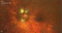
Morning Glory Disc Anomaly | Bashaer Aldhahwani, MD; Carlos Ernesto Mendoza Santiesteban, MD | A colored fundus photo of a patient with morning glory anomaly. Morning glory anomaly is a rare congenital malformation of the optic nerve. The morning glory disc anomaly can be seen with transsphenoidal basal encephalocele. It is known as morning glory syndrome when it is associated with systemic s... | Optic Disc Anomaly |
| 90 |
 |
Curtain Sign (Enhanced Ptosis) | Bashaer Aldhahwani, MD; Hong Jiang, MD, PhD | This is a 78-year-old male patient who presented with diplopia, right eyelid ptosis, and ophthalmoplegia. He had severe ptosis OD and pseudo-proptosis (lid retraction) OS at baseline, but when the right eyelid was manually elevated, there was marked enhanced ptosis of the left eyelid (Video). He was... | Myasthenia GravIs; Clinical Signs |
| 91 |
 |
Curtain Sign (Enhanced Ptosis) - Associated Image 1 | Bashaer Aldhahwani, MD; Hong Jiang, MD, PhD | This is a 78-year-old male patient who presented with diplopia, right eyelid ptosis, and ophthalmoplegia. He had severe ptosis OD and pseudo-proptosis (lid retraction) OS at baseline, but when the right eyelid was manually elevated, there was marked enhanced ptosis of the left eyelid (Video). He was... | Myasthenia GravIs; Clinical Signs |
| 92 |
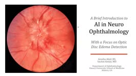 |
A Brief Introduction to AI in Neuro-ophthalmology | Areeba Abid, BS; Sachin Kedar, MD | In this video, we describe the basics of artificial intelligence and machine learning as applicable to clinical neuro-ophthalmologists. We use the example from a recent publication, where AI software was used to detect optic disc edema in fundus images. | Artificial Intelligence; Machine Learning |
| 93 |
 |
Optic Nerve Hypoplasia (ONH) - Double Ring Sign | Bashaer Aldhahwani, MD; Joshua Pasol, MD | Optic nerve hypoplasia (ONH) is characterized by a decreased number of optic nerve axons. It can present unilaterally or bilaterally, Isolated or associated with midline cerebral structural defects, such as septum pellucidum absence, agenesis of corpus callosum, cerebral hemisphere abnormalities, or... | Optic Nerve Hypoplasia (ONH) |
| 94 |
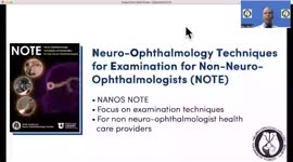 |
Introduction to NANOS NOTE | Karl C. Golnik, MD | Introduction to NANOS NOTE, a resource for non-neuro-ophthalmologists describing common examination techniques. | Neuro-Ophthalmology Examination Techniques |
| 95 |
 |
Myelinated Retinal Nerve Fiber Layer | Bashaer Aldhahwani, MD; Hong Jiang, MD, PhD | A 78 YOF with no visual symptoms has an incidental finding of yellow-white well-demarcated patches with ragged borders at the peripapillary area of her left eye (see the fundus photo). | Myelinated Retinal Nerve Fiber Layer |
| 96 |
 |
Ocular Neuromyotonia Video | Bashaer Aldhahwani, MD; Joshua Pasol, MD | A video demonstrates ocular neuromyotonia in the left eye of a patient with a history of cranial radiation of parasellar mass. Ocular neuromyotonia (ONM) is a rare ocular motor disorder characterized by intermittent, tonic spasms of one or more of the extraocular muscles, resulting in strabismus and... | Ocular Neuromyotonia |
| 97 |
 |
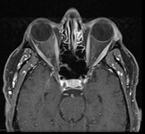
Indirect Carotid Cavernous Fistula | Edsel Ing, MD, PhD, FRCSC | A 67-year-old woman had delayed initial diagnosis of her right low flow carotid cavernous fistula (CCF) during the coronavirus disease (COVID-19) pandemic due to difficulty detecting ocular signs via online virtual examinations. Her right eye conjunctival erythema and proptosis with medial rectus en... | Carotid Cavernous Fistula; Misdiagnosis; Radiology |
| 98 |
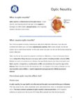 |
Optic Neuritis | NANOS | In the most common form of optic neuritis, the optic nerve has been attacked by the body's overactive immune system and results in decreased vision. | Optic Neuritis; Patient Brochure |
| 99 |
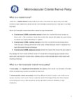 |
Microvascular Cranial Nerve Palsy | NANOS | Microvascular cranial nerve palsy is one of the most common causes of double vision in the older poulation. They are often referred to as "diabetic" palsies. They will resolve without leaving any double vision. | Microvascular Cranial Nerve Palsy; Patient Brochure |
| 100 |
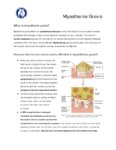 |
Myasthenia Gravis | NANOS | This is an autoimmune condition where the body's immune system has damaged receptors on your muscles and can result in double vision or drooping lid. | Myasthenia Gravis; Patient Brochure |