A collection of videos relating to the diagnosis and treatment of eye movement disorders. This collection includes many demonstrations of examination techniques.
Dan Gold, D.O., Associate Professor of Neurology, Ophthalmology, Neurosurgery, Otolaryngology - Head & Neck Surgery, Emergency Medicine, and Medicine, The Johns Hopkins School of Medicine.
A collection of videos relating to the diagnosis and treatment of eye movement disorders.
NOVEL: https://novel.utah.edu/
TO
Filters: Collection: "ehsl_novel_gold"
| Title | Description | Type | ||
|---|---|---|---|---|
| 76 |
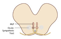 |
Figure 69: Vascular Distribution and Anatomy (Including 4th Nerve) of the Caudal Midbrain (Supplement) | Image | |
| 77 |
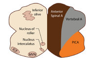 |
Figure 80: Vascular Distribution and Anatomy Relevant to the Medial Medullary Syndrome (Supplement) | Image | |
| 78 |
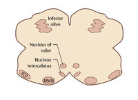 |
Figure 80: Vascular Distribution and Anatomy Relevant to the Medial Medullary Syndrome (Supplement) | Image | |
| 79 |
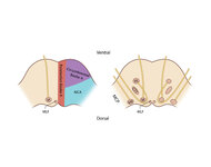 |
Figure 61: Vascular Distribution and Anatomy (Including 6th, 7th, 8th Nerves, MLF) of the Pons | In this axial section of the pons, the proximity of the 7th (VII) and 8th (VIII) fascicles can be appreciated, and a lateral inferior pontine syndrome (anterior inferior cerebellar artery, AICA territory), which could involve both of these fascicles, could cause acute prolonged vertigo accompanied b... | Image |
| 80 |
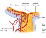 |
Figure 27: Vascular Supply of the Optic Nerve Head, Choroid and Retina | The ophthalmic artery is a branch of the internal carotid artery, which in turn, supplies the posterior ciliary (to choroid and outer retina) and central retinal (to inner retina) arteries. The central retinal artery (CRA) enters the optic nerve about 1 cm posterior to the globe, and an embolus may ... | Image |
| 81 |
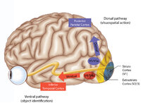 |
Figure 43: How the Brain Makes Sense of What It Sees - The Dorsal and Ventral Visual Pathways, and a 3 Tiered Approach to Vision | 1) Ventral ("what") stream - this begins with the ‘P' retinal ganglion cells à parvocellular layers of the lateral geniculate nucleus (LGN, 3-6) à V1/striate cortex (in blue) à V4/V4a (fusiform and lingual gyri) à occipitotemporal regions. 2) Dorsal ("where") stream - this begins with the ‘M... | Image |
| 82 |
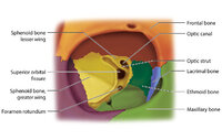 |
Figure 17: Bony Structures Relevant to the Orbit | The frontal, sphenoid, maxillary, ethmoid, and lacrimal bones make up the orbit. Structures passing through the optic canal include the optic nerve, oculosympathetic tract and ophthalmic artery. Structures passing through the superior orbital fissure include the superior ophthalmic vein, cranial ner... | Image |
| 83 |
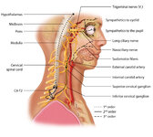 |
Figure 1: Oculosympathetic Pathway for Pupillary Dilation | The oculosympathetic tract is an uncrossed pathway that begins in the hypothalamus, with fibers descending in the brainstem (1st order, commonly affected in a lateral medullary syndrome), synapsing in the lower cervical/upper thoracic spinal cord (interomediolateral cell columns of C8-T2, also refer... | Image |
| 84 |
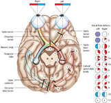 |
Figure 24: Typical Visual Field Defects Associated with Discrete Lesions Along the Visual Pathways | Specific monocular or binocular visual field defects can be highly localizing when the neuroanatomy of the visual pathways is understood. The temporal visual field corresponds to the nasal retina, while the nasal visual field corresponds to the temporal retina. 1) Left optic nerve lesion - while an ... | Image |
| 85 |
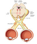 |
Figure 2: Parasympathetic Pathway for Pupillary Constriction | A bright light is shone in one eye, light enters the pupil and hyperpolarizes retinal photoreceptors which activates retinal ganglion cells. These signals propagate along the optic nerves, chiasm, optic tracts, and fibers responsible for the light reflex then synapse in the dorsal midbrain (prior to... | Image |
| 86 |
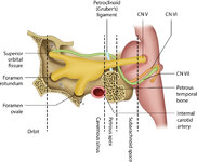 |
Figure 46: The Course of the 6th (VI) Nerve | The sixth nucleus is located dorsally, adjacent to the 4th ventricle, in the lower pons. The genu of the facial (7th) nerve wraps around the 6th nucleus, creating the facial colliculus, which bulges into the 4th ventricle. After the 6th nerve leaves the pons, it follows a vertical course along the c... | Image |
| 87 |
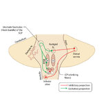 |
Figure 50: Anatomy and Physiology of the Saccadic Pathways | When a saccade is desired (or reflexively triggered), signals project from the saccade-related cortical eye fields to the superior colliculus, which serves to integrate and relay commands to the saccade generating brainstem circuitry. The inferior cerebellar peduncle (ICP) carries climbing fibers to... | Image |
| 88 |
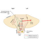 |
Figure 51: Lateral Medullary Lesion Causing Saccadic Dysmetria | A lesion of the left lateral medulla and inferior cerebellar peduncle (ICP) will cause decreased climbing fiber inhibition of the left dorsal vermis causing simple-spike (inhibitory) discharge of Purkinje cells to increase. Increased Purkinje cell firing leads to increased inhibition of the ipsilate... | Image |
| 89 |
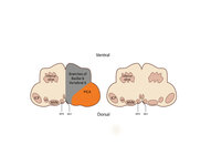 |
Figure 53: Vascular Distribution and Anatomy Relevant to the Lateral Medullary (Wallenberg) Syndrome | This axial section of the medulla highlights those structures that, when damaged, are responsible for the vestibular and ocular motor features of the Wallenberg syndrome. The nucleus prepositus hypoglossi (NPH) and medial vestibular nucleus (MVN) complex is important for horizontal gaze-holding (neu... | Image |
| 90 |
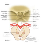 |
Figure 64: The Course of the 3rd (III) Nerve | The 3rd nucleus lies at the ventral border of the periaqueductal gray matter, at the level of the superior colliculus. In between the two nuclei is the midline central caudal nucleus (CCN), which innervates bilateral levator palpebrae muscles (explaining how a unilateral nuclear 3rd can cause bilate... | Image |
| 91 |
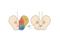 |
Figure 65: Vascular Distribution and Anatomy (Including 3rd Nerve) of the Rostral Midbrain | In this axial section of the midbrain at the level of the superior colliculus, the paired 3rd nuclei are located ventral to the periaqueductal grey, and the midline central caudal nucleus (CCN) is located in between. The fascicles that exit the IIIrd nuclei carry the fibers destined to innervate the... | Image |
| 92 |
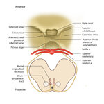 |
Figure 68: The Course of the 4th (IV) Nerve | The 4th nucleus lies at the ventral border of the periaqueductal gray matter, at the level of the inferior colliculus. The fascicles exit the nucleus dorsally and decussate at the anterior medullary velum (anterior floor of the fourth ventricle), and then exit the brainstem dorsally. The peripheral ... | Image |
| 93 |
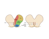 |
Figure 69: Vascular Distribution and Anatomy (Including 4th Nerve) of the Caudal Midbrain | In this axial section of the midbrain at the level of the inferior colliculus, the 4th nuclei are located ventral to the periaqueductal grey, dorsal to the medial longitudinal fasciculus (MLF) and medial to the oculosympathetic tract. Fascicles exit the nucleus dorsally and decussate at the anterior... | Image |
| 94 |
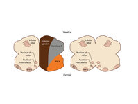 |
Figure 80: Vascular Distribution and Anatomy Relevant to the Medial Medullary Syndrome | This axial section of the medulla highlights those structures that, when damaged, are often responsible for spontaneous upbeat nystagmus (UBN). The nucleus of Roller and nucleus intercalatus normally have an inhibitory influence over the cerebellar flocculus, and when there is a lesion of Roller/int... | Image |
| 95 |
 |
Ocular Dipping and Ping-pong Gaze Due to Bi-hemispheric Strokes | This is a 51-year-old man presenting with hypertensive left thalamic intracerebral hemorrhage and intraventricular hemorrhage, with course complicated by multifocal supratentorial ischemic strokes. He developed abnormal movements characterized by slow, conjugate, horizontal deviations, consistent wi... | Image/MovingImage |
| 96 |
 |
Centripetal Nystagmus Example | A 68-year-old female reported a 2-year history of progressive gait imbalance, falls, dizziness and vertical oscillopsia. She described that dizziness and oscillopsia were worst when looking down. There was no family history of ataxia. Composite gaze with fixation was recorded with video-oculography ... | Image/MovingImage |
| 97 |
 |
CANVAS (Cerebellar Ataxia, Neuropathy, and Vestibular Areflexia Syndrome) Video Head Impulse Test (vHIT) Figure | CANVAS (Cerebellar Ataxia, Neuropathy, and Vestibular Areflexia Syndrome) is a genetic condition consisting of slowly progressive late-onset ataxia, bilateral vestibulopathy, sensory neuropathy, chronic cough, and autonomic dysfunction. While the term vestibular areflexia typically implies bilateral... | Image |
| 98 |
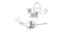 |
Atypical PC BPPV Variant Figures | Figure 1: Atypical posterior canal BPPV variants The labyrinth consists of the cochlea (C), two otolithic organs including utricle (U) and saccule (S), and three semicircular canals including anterior canal (AC), horizontal canal (HC), and posterior canal (PC). A. If otoconia are located within the ... | Image |
| 99 |
 |
Assessing Utricle Pathway Function and the Effects of Convergence on Nystagmus in Acute Vestibular Neuritis | A 35-year-old woman presented a few days after the onset of room-spinning vertigo. She denied diplopia, dysarthria, dysphagia, dysphonia, incoordination, numbness, and weakness. On examination, she had subtle spontaneous right-beat nystagmus (RBN). This nystagmus increased in amplitude and frequency... | Image/MovingImage |
| 100 |
 |
Upbeat Transitioning to Downbeat Nystagmus in Wernicke's Encephalopathy | This is a 30-year-old man with a history of alcohol abuse who presented to the hospital with inability to walk after several weeks of heavy drinking and malnutrition. While in the hospital, he noted that when he would look straight ahead, everything he saw would appear to be bouncing up and down - a... | Image/MovingImage |