AAO-NANOS Neuro-Ophthalmology Clinical Collection: Derived from the AAO-NANOS Clinical Neuro-Ophthalmology collection produced on CD. The images are of selected cases from the NANOS teaching slide exchange, and the CD was produced under the direction of Larry Frohman, MD and Andrew Lee, MD.
The American Academy of Ophthalmology (AAO); The North American Neuro-Ophthalmology Association (NANOS).
NOVEL: https://novel.utah.edu/
TO
Filters: Collection: "ehsl_novel_aao_nanos"
| Title | Creator | Description | ||
|---|---|---|---|---|
| 76 |
 |
Ocular Manifestations of Systemic Disorders | Rosa A. Tang, MD | Myasthenia gravis should be considered in any patient with painless, pupil-spared, nonapoptotic ophthalmoplegia. It may mimic any ophthalmoparesis. Involvement of the medical rectus may result in a pseudointernuclear ophthalmoplegia. Pair with 96_24 and 96_25. |
| 77 |
 |
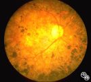
Optic Nerve Drusen, Late Fluorescein Angiogram | Anthony C. Arnold, MD | Images 92_64 and 92_67 demonstrate the characteristics of optic disc drusen on flourescein angiography. This image displays the nodular staining of the drusen without leakage. Pair with 92_63 and 92_64. |
| 78 |
 |
Motility Disturbances | Don Bienfang, MD | This patient displays a posttraumatic left fourth nerve palsy sustained after having struck her head on the dashboard. |
| 79 |
 |
Motility Disturbances | Don Bienfang, MD | This patient displays a posttraumatic left fourth nerve palsy sustained after having struck her head on the dashboard. |
| 80 |
 |
Motility Disturbances | Don Bienfang, MD | This patient displays a posttraumatic left fourth nerve palsy sustained after having struck her head on the dashboard. |
| 81 |
 |
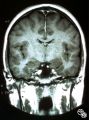
Systemic Disorders With Optic Nerve and Retinal Findings | Larry P. Frohman, MD | A 35-year-old African-American woman had gradual bilateral painless visual loss over 3 months. When initially seen, the visual acuities were HM OD, NLP OS. The MRI showed diffused enhancement of the optic nerves and lacrimal glands. The evaluation strongly suggested sarcoidosis, with elevated angiot... |
| 82 |
 |
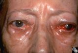
Ocular Manifestations of Systemic Disorders | Larry P. Frohman, MD | A 17-year-old girl had undergone multiple superficial biopsies of the orbit for what was felt to be refractory orbital pseudotumor. Initial evaluation revealed the saddle-nose deformity, which the patient confirmed was acquired. More extensive biopsy was consistent with lymphomatoid granulomatosis. ... |
| 83 |
 |
Ocular Manifestations of Congenital/Inherited Diseases | Jacqueline A. Leavitt, MD | This 22-year-old woman has neurofibromatosis, type 2. Acuity, color plates, pupillary responses, slit-lamp examination, IOP, fields, and funduscopy are all normal. There is a 3 mm proptosis OS. The patient has recently undergone gamma knife for the acoustic tumor, and she has residual facial nerve p... |
| 84 |
 |
Motility Disturbances | Rosa A. Tang, MD | Traumatic damage to the third cranial nerve may result in aberrant regeneration of fibers that innervate the eyelid, pupil, or extraocular muscles. For instance, there may be lid retraction in attempted downgaze. Any combination of aberrant activation of third nerve-innervated structures may occur, ... |
| 85 |
 |
Isolated Congenital Optic Disc Anomalies | Thomas R. Wolf, MD | An optic pit is a small defect in the optic disc that may be asymptomatic in isolation. Patients may develop an associated serous detachment of the macula. The condition is usually unilateral but may be bilateral. A fluorescein angiogram may demonstrate the serous detachment, and laser photocoagulat... |
| 86 |
 |
Ocular Manifestations of Systemic Disorders | Rosa A. Tang, MD | Thyroid eye disease may cause proptosis and extraocular muscle enlargement that may be seen on orbital imaging studies. In general, coronal images allow the best visualization of the extraocular muscle enlargement. Pair with 94_45 and 94_46. |
| 87 |
 |
Orbital Tumors | Mitchell J. Wolin, MD | Cavernous hemangiomas of the orbit usually result in painless orbital signs such as proptosis or visual loss. Orbital imaging of the lesion, which usually is a well-defined orbital mass, is demonstrated in this study. The lesion is benign and usually occurs in young to middle-aged adults. Surgical e... |
| 88 |
 |
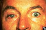
Ocular Manifestations of Systemic Disorders | Rosa A. Tang, MD | Myasthenia gravis should be considered in any patient with painless, pupil-spared, nonapoptotic ophthalmoplegia. It may mimic any ophthalmoparesis. Involvement of the medical rectus may result in a pseudointernuclear ophthalmoplegia. Pair with 96_23 and 96_25. |
| 89 |
 |
Ocular Manifestations of Systemic Disorders | Rosa A. Tang, MD | Myasthenia gravis should be considered in any patient with painless, pupil-spared, nonapoptotic ophthalmoplegia. It may mimic any ophthalmoparesis. Involvement of the medical rectus may result in a pseudointernuclear ophthalmoplegia. Pair with 96_23 and 96_24. |
| 90 |
 |
Ocular Manifestations of Congenital/Inherited Diseases | Mitchell J. Wolin, MD | Patients with olivopontocerebellar atrophy may exhibit signs of ocular motor deficits, such as ocular motor apraxia or cerebellar eye signs, and peripheral pigmentary retinopathy and optic atrophy. Pair with 94_55. |
| 91 |
 |
Ocular Manifestations of Congenital/Inherited Diseases | Mitchell J. Wolin, MD | Patients with olivopontocerebellar atrophy may exhibit signs of ocular motor deficits, such as ocular motor apraxia or cerebellar eye signs, and peripheral pigmentary retinopathy and optic atrophy. Pair with 94_54. |
| 92 |
 |
Isolated Optic Neuritis/Neuropathy | Rosa A. Tang, MD | Papilledema may produce visual loss due to chronic atrophic papilledema, secondary macular hemorrhage, exudate or edema, secondary ischemic optic neuropathy, or secondary subretinal neovascular membrane formation. |
| 93 |
 |
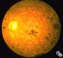
Optic Disc Drusen With Autofluorescence | Thomas R. Wolf, MD | This photograph of optic disc drusen demonstrates autoflourescence with flourescein barrier filters in place. Imaging: flourescein barrier filters. |
| 94 |
 |
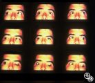
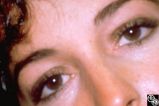
Migraine Syndrome | Mitchell J. Wolin, MD | The image shows a patient with cluster headache and eye displaying Horner's syndrome. |
| 95 |
 |
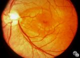
Isolated Congenital Optic Disc Anomalies | Thomas R. Wolf, MD | Benign tumors of blood vessels (hemangiomas) may occur on the optic nerve and may mimic optic disc edema. Disease/Diagnosis: Optic Nerve Hemangioma. |
| 96 |
 |
Motility Disturbances | Don Bienfang, MD | This patient displays a posttraumatic left fourth nerve palsy sustained after having struck her head on the dashboard. |
| 97 |
 |
Isolated Optic Neuritis/Neuropathy | Rosa A. Tang, MD | Papilledema in pseudotumor cerebri may result in adjacent choroidal or retinal folds. |
| 98 |
 |
Optic Tract Syndrome Due to Carotid Artery Dolichoectasia | Larry P. Frohman, MD | This 43-year-old man was referred for evaluation of 6 months of visual loss OU. In retrospect, he had noticed increasing difficulty with his tennis game dating back over 3 years, as balls would pass him unexpectedly when hit to his backhand (left) side. The patient did not think this was progressive... |
| 99 |
 |
Optic Tract Syndrome Due to Carotid Artery Dolichoectasia | Larry P. Frohman, MD | This 43-year-old man was referred for evaluation of 6 months of visual loss OU. In retrospect, he had noticed increasing difficulty with his tennis game dating back over 3 years, as balls would pass him unexpectedly when hit to his backhand (left) side. The patient did not think this was progressive... |
| 100 |
 |
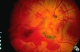
Isolated Congenital Optic Disc Anomalies | Rosa A. Tang, MD | This patient has optic disc drusen and evidence of a superimposed optic neuropathy, including loss of visual field, an ipsilateral afferent pupillary defect, and optic atrophy. Although optic disc drusen typically causes visual field loss without visual acuity loss superimposed, ischemic optic neuro... |