The Health Education Assets Library (HEAL) is a collection of over 22,000 freely available digital materials for health sciences education. The collection is now housed at the University of Utah J. Willard Marriott Digital Library.
TO
Filters: Collection: "ehsl_heal" Format: image
| Title | Description | Subject | Collection | ||
|---|---|---|---|---|---|
| 76 |
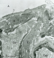 |
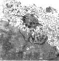
Elastin in alveolar tip in lung tissue (human, adult) | Immuno-electron microscopy (embedded in Lowicryl HM 20). The amorph elastin (E) appears pale after embedding in this type of resin, but is antibody-labeled with electron-dense gold particles of 10 nm. (Col) indicate bundles of collagen fibers which are intermingled with swollen cytoplasmic extension... | Alveolar cell type I; Myofibroblast; Immuno-electron microscopy; Immuno-staining | Poja Histology Collection - Respiratory System Subset |
| 77 |
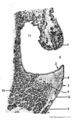 |
Scheme survey frontal section of larynx (human) | Stain: Azan. A pseudostratified epithelium covers the mucosa of the laryngeal cavity and vestibulum. At the vocal fold edge (3) the epithelium appears to be nonkeratinizing squamous. The connective tissue is rigidly attached to this edge and merges into the vocal ligament (dense elastic) and vocal m... | Vocal ligament; Laryngeal glands; Ventricular fold | Poja Histology Collection - Respiratory System Subset |
| 78 |
 |
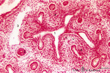
Pseudoglandular period of developing lung (human, embryo, low magnification) | Stain: Hematoxylin and eosin. The two future lung lobes contain many cross-sectioned future bronchial tubes (1). Note in these epithelial cells the apical position of the nuclei with light-stained basal parts (glycogen). The surrounding mesenchyme becomes more condensed (↓) around the epithelium a... | Lung development; Visceral pleura; Pseudoglandular period; Bronchial tubes; Mesenchyme | Poja Histology Collection - Respiratory System Subset |
| 79 |
 |
Terminal sac period of developing lung (human, fetus, low magnification) | Stain: Hematoxylin and eosin. At the right cross-sections of a large bronchus (1) with cartilage rings (2). Close to them a pulmonary artery (3), and a bronchiolus (4) without cartilage. (5) indicates a respiratory bronchiolus. Most alveoli are not inflated, the numerous dilated spaces (6) are saccu... | Lung development ; Terminal sac period; Respiratory bronchioli | Poja Histology Collection - Respiratory System Subset |
| 80 |
 |
Olfactory vesicle (bulb) in the nose (gerbil) | DUPLICATE RECORD - FOR DELETION Electron microscopy. A distinct olfactory bulb (1, vesicle) with horizontally extended cross-sectioned cilia (*) and basal bodies (↑) is surrounded by numerous microvilli and free olfactory cilia. Close to the first bulb a second one is about to protrude above th... | Olfactory epithelium; Olfactory vesicle | Poja Histology Collection - Respiratory System Subset |
| 81 |
 |
Ventricular fold (plica ventricularis) (human) | Stain: Azan. On top pseudostratified epithelium (1). Lamina submucosa with predominantly mucous glands (3), fat cells (4) and blood vessels. The lamina propria also contains lymphoid follicles (2) with a germinal center (immune protection). | Laryngeal glands; Ventricular fold; Seromucous glands | Poja Histology Collection - Respiratory System Subset |
| 82 |
 |
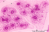
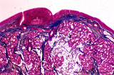
Survey of a centrilobular lung emphysema: section of an lobule (human, adult) | Stain: Hematoxylin and eosin. The enlargement of large areas of the air spaces (X) is evident due to destruction of the walls of alveoli throughout the lobule. | Poja Histology Collection - Respiratory System Subset | |
| 83 |
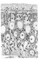 |
Olfactory epithelium in the nasal cavity (mammals) | Scheme electron microscopy. Four olfactory bulbs (1, vesicles) with radially sprouting cilia are present at the surface of the epithelium. Their slender cell bodies (2, bipolar neurons) are flanked by sustentacular (supporting) broad cells (3) whose apices are filled with well developed organelles, ... | Pseudostratified epithelium; Olfactory epithelium; Basal cells; Olfactory vesicle; Bipolar neuron | Poja Histology Collection - Respiratory System Subset |
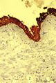
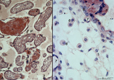
| 84 |
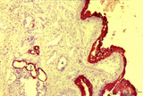 |
Keratin staining of bronchus epithelium (human, adult) | Stain: Anti-keratin-5-7-14-19 (OVTL 12/5 monoclonal antibody with immunoperoxidase staining on frozen sections). The pseudostratified epithelium (1) is stained keratin-positively (brown product) as well as the epithelial derivates such as the bronchial glands (2) and their excretory ducts (3). Other... | Bronchial epithelium; Keratin antibodies; Tumor diagnosis | Poja Histology Collection - Respiratory System Subset |
| 85 |
 |
Keratin staining of bronchus epithelium (human, adult) | Stain: Anti-keratin-5-7-14-19 (OVTL 12/5 monoclonal antibody with immunoperoxidase staining on frozen sedctions). The pseudostratified epithelium (1) reacts positively (brown-red product), the nuclei of the basal cells are well visible (arrows). Other structures react negatively (blue stain) such as... | Tumor diagnosis; Bronchial epithelium; Lung parenchym; Keratin antibodies | Poja Histology Collection - Respiratory System Subset |
| 86 |
 |
Longitudinal section of trachea (human, adult) | Stain: Azan. On top: the epithelium (1) is pseudostratified with cilia and goblet cells on a distinct basement membrane (↓). There is almost no demarcation (at this magnification) between lamina propria and submucosa due to the dense blue (2) staining of elastic fibers reinforced by collagen fiber... | Pseudostratified epithelium ; Tracheal glands; Fibroelastic membrane | Poja Histology Collection - Respiratory System Subset |
| 87 |
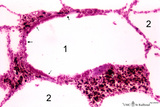 |
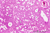
Respiratory bronchiolus (mouse) | Stain: Hematoxylin and eosin. The lumen (1) shows an irregular lining and is covered by low columnar-cuboidal cells (↓), locally disrupted by a few areas with thin alveolar epithelium as found in the surrounding alveoli (2). Thick arrows point to a row of light-stained Clara cells. At (*) discont... | Respiratory bronchiolus; Columnar epithelium; Cuboidal epithelium; Clara cells; Alveolar epithelium | Poja Histology Collection - Respiratory System Subset |
| 88 |
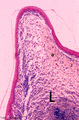 |
Vocal cord (plica vocalis) (human, glottis) | Stain: Azan. The true vocal cord consist of the vocal ligament (L) covered by stratified squamous epithelium and the striated vocal muscle (medial part of the thyreoarythenoid muscle, not depicted in this picture). At the tip of the vocal cord the stratified epithelium is distinct and the lamina pro... | Vocal ligament | Poja Histology Collection - Respiratory System Subset |
| 89 |
 |
Pseudoglandular - canalicular period of developing lung (human, fetus) | Stain: Azan. Terminal budding and elongation of the future bronchial tree: branching of a bronchial tubes (1) within an islet where the mesenchyme condenses (4). A large blood vessel (2) as well as a lymph vessel (3) are recognizable. Note in the mesenchyme formation of blood capillaries in the vici... | Lung development; Pseudoglandular period ; Canalicular period; Mesenchyme | Poja Histology Collection - Respiratory System Subset |
| 90 |
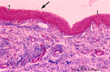 |
Transition surface of vocal cord into laryngeal surface (human, subglottis, higher magnification) | Stain:Stain: Azan. Transition (↓) from stratified squamous epithelium (1) into pseudostratified epithelium (2) in the subglottis region. Blood vessels and the cellularity of the proper lamina is evident. | Olfactory epithelium; Subglottis; Stratified epithelium; Pseudostratified epithelium | Poja Histology Collection - Respiratory System Subset |
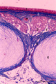
| 91 |
 |
Submucosa of trachea (human, adult) | Stain: Goldner trichrome. Mixed tracheal glands (1) of the submucosa (connective tissue light green) and bundles of smooth muscle fibers (2) close to the fibroelastic membrane between the cartilage edges. | Seromucous gland; Tracheal glands | Poja Histology Collection - Respiratory System Subset |
| 92 |
 |
Tertiary villi (human placenta, late midpregnancy) | : (A) Left: stain: Hematoxylin - azophloxine. (B) Right: electron microscopy (low magnification). At the left (A) multinucleated syncytiotrophoblast cells (STCs, 1) and vaguely cytotrophoblast cells (CTCs, 2) cover the loose fetal stroma (4). A large and a small arteriole (3) are embedded with stro... | placenta; tertiary villi; placental barrier; Hofbauer cell; vasculosyncytial membrane; syncytiotrophoblast | Poja Histology Collection - Placenta |
| 93 |
 |
Survey of implanted blastocyst (human, ca. 3 weeks-old embryo) and detail of advanced stage of decidua cells (human, mid pregnancy) | Stain: Hematoxylin-eosin (A, left) and Azan (B, right). (A) Left: section through a slightly damaged implantation site shows at (1) the uterine lumen and (2) the decidua capsularis while at (3) several uterine glands are visible. At arrow (4) the amniotic cavity, (5) points to embryonic shield and... | placenta; uterus; syncytiotrophoblast; implantation; blastocyst | Poja Histology Collection - Placenta |
| 94 |
 |
Tertiary villi (human placenta, midpregnancy) | Stain: Hematoxylin - azophloxine. (A) Left a variety of tertiary villi and reddish-stained fibrinoid depositions (1). At (2): the embryonic connective tissue starts to fibrinize. Note syncytiotrophoblast knots (3) or sprouts of several villi .These knots might represent aggregations of degenerati... | placenta; fibrinoid; cytotrophoblast; syncytiotrophoblast | Poja Histology Collection - Placenta |
| 95 |
 |
Syncytiotrophoblast cells of tertiary villi (human placenta, midpregnancy) | (A) Left: stain: Immunoperoxidase staining with diaminobenzidin (DAB) and human anti-chorionic gonadotrophin antibody (hCG-DAKO 231), counterstained with hematoxylin. (B) Right: electron microscopy. (A) At the left the exclusive reactivity (dark brown) in the cytoplasma of the multinucleated syn... | Syncytiotrophoblast; placenta ; chorionic villi; HCG; immunohistochemistry; electron microscopy | Poja Histology Collection - Placenta |
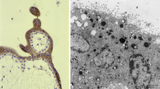
| 96 |
 |
Electron microscopy of tertiary villus (human placenta, early pregnancy) | At the left (A), part of a tertiary (terminal) villus with the multinucleated syncytiotrophoblast cell (1) (STC) at the top. The apex shows protrusions and extensive microvilli of varying sizes (brushborder). Nuclei (1a) are sectioned at different levels and the cytoplasm contains abundant organelle... | placenta; chorionic villi; placental barrier; syncytiotrophoblast; electron microscopy | Poja Histology Collection - Placenta |
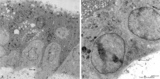
| 97 |
 |
Electron microscopy of syncytiotrophoblast cells in tertiary villus (human placenta, midpregnancy) | The left photograph (A) shows the apical cytoplasm of a syncytiotrophoblast cell (STC, nucleus). The free cell surface displays small protrusions and a characteristic pattern of differently shaped microvilli; pinocytotic invaginations and a single macropinocytotic vacuole. Note the apical localized ... | placenta; tertiary villi; syncytiotrophoblast; electron microscopy | Poja Histology Collection - Placenta |
| 98 |
 |
Survey of the chorionic plate and intervillous space (human placenta, full-term) | Stain: Perjodic acid-Schiff reaction (PAS). At the top the chorionic plate (1) with cross-sections of umbilical vessels (2) At (3) the folded amnion covering the chorionic plate. Ramifications of thicker stem villi demonstrate free-floating terminal villi (tertiary). At several places reddish-st... | placenta; tertiary villi; decidua; chorionic plate | Poja Histology Collection - Placenta |
| 99 |
 |
Stem villus (human placenta, midpregnancy) | Stain: Trichrome (Goldner). At the top the fetal site with part of the chorionic plate (1) with a thick stem villus (2) ramifying in two smaller villi with large blood vessels (3). The chorionic plate consists of a thick fibrous connective layer (green). Towards the intervillous space it is covere... | placenta; chorionic villi; stem villus; chorion plate | Poja Histology Collection - Placenta |
| 100 |
 |
Spiral artery in endometrium (human, midpregnancy) | Stain: Hematoxylin-eosin. (A) Emerging into partly damaged intervillous space (1), filled with hemorrhage and fibrin (2). (3) uterine vein and (4) smooth muscle cell bundles at the endometrial-myometrial junction. (B) Lumen with hypertrophic endothelium at higher magnification (5). Note as an unu... | placenta; trophoblast; spiral artery; endometrium | Poja Histology Collection - Placenta |