The Health Education Assets Library (HEAL) is a collection of over 22,000 freely available digital materials for health sciences education. The collection is now housed at the University of Utah J. Willard Marriott Digital Library.
TO
| Title | Description | Subject | Collection | ||
|---|---|---|---|---|---|
| 76 |
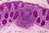 |
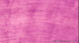
Colon ('gut-associated lymphatic tissue' or GALT) (human) | Stain: Hematoxylin-PAS. Solitary lymphatic nodule in colon (see also Digestive System: Colon) A large amount of non-encapsulated diffuse lymphatic tissue or mucosa-associated lymphatic tissue (MALT) is located in the subepithelial lamina propria of the colon and called gut-associated lymphatic tiss... | GALT; follicle; lymphoid tissue | Poja Histology Collection - Lymphatic Tissues and Organs Subset |
| 77 |
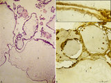 |
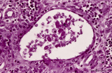
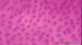
Complete hydatidiform mole (human) | Stain: (A) Hematoxylin-eosin. (B+C) Immunoperoxidase staining with diaminobenzidin (DAB) and hematoxylin counterstaining for keratin 7 (OVTL 12-30 antibody). (A)Trophoblast cells surround the dilated chorionic villi (1), with centrally large amounts of edematous mesenchymal stroma with almost no b... | trophoblast; complete hydatidiform mole; placenta; villus; immunohistochemistry; keratin-7 | Poja Histology Collection - Placenta |
| 78 |
 |
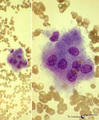
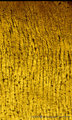
Contamination with osteoblasts of a bone marrow puncture (human) | Stain: May-Grnwald-Giemsa (MGG). In bone marrow smears from a sternum puncture sometimes osteoblasts are found. Osteoblasts may occur in groups, are oval with a usually eccentric, relatively small nucleus. The chromatin is often coarse with 1-3 nucleoli. The cytoplasm is mostly light blue and may co... | Poja Histology Collection - Blood & Bone Marrow Subset | |
| 79 |
 |
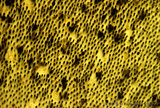
Contamination with osteoclasts of a bone marrow puncture (human) | Stain: May-Grnwald-Giemsa (MGG). In the procedure for preparing a bone marrow smear from a sternum puncture osteoclasts are frequently found in the bone marrow smears. The osteoclasts are multinucleated giant cells for the destruction of bone in the process of remodelling and growth. The cytoplasm m... | Poja Histology Collection - Blood & Bone Marrow Subset | |
| 80 |
 |
Contamination with squamous cell cells in peripheral blood smear (A) and in a bone marrow smear (B) (human) | Stain: May-Grnwald-Giemsa (MGG). In blood and bone marrow smears incidentally squamous epithelial cells from the skin tissues are found. (A-1) is an epithelial cell from the keratinized stratified (upper) epidermis; (A-2) a young neutrophil with a few vacuoles in the cytoplasm (probably degeneration... | Poja Histology Collection - Blood & Bone Marrow Subset | |
| 81 |
 |
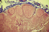
Corpuscles in thymus (human, puberty) | Stain: Hematoxylin. A: Several thymic (Hassall's) corpuscles (*) of varying sizes within the medulla (1). The Hassall bodies are surrounded by recognizable flattened cells showing keratohyalin granules. The outer shell consists of more layers of close packed concentrically arranged cells with light-... | thymic corpuscle (Hassalls); epithelioreticular cell (ERC); thymus medulla; lymphoid tissue | Poja Histology Collection - Lymphatic Tissues and Organs Subset |
| 82 |
 |
Cortex of lymph node (human) | Stain: Azan. The cortex of the lymph node comprises the (secondary) follicles with germinal centres (1) and corona or mantle zone (2), all filled with B lymphocytes. The paracortical area (3) below the follicles largely comprises T cells. Afferent lymph vessels enter via the capsule (6) into the m... | cortex; paracortex | Poja Histology Collection - Lymphatic Tissues and Organs Subset |
| 83 |
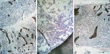 |
Crista biopsies of normal, hypoplastic and malignant bone marrow (human) | Stain: Hematoxylin and eosin. The composition of three slides shows a normal bone marrow section (A), an aplastic bone marrow (B), and a malignant lymphoma in bone marrow (C). (1) Cristae. (2) Fat tissue or fat cells. (3) Erythrocytes and (4) erythroblasts with dark nuclei. | Poja Histology Collection - Blood & Bone Marrow Subset | |
| 84 |
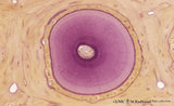 |
Cross section of tooth in alveolar bone - cat; low magnification | Stain: Picric acid and hematoxylin. From left to right: alveolar bone tissue with osteons; periodontal ligament with blood vessels; acellular cementum (dark purple rim); dentin with (purple) radiair arrangement of dentinal tubules; fine incremental (imbrication) lines of von Ebner run at right angl... | oral cavity; incremental lines; alveolar bone | Poja Histology Collection - Oral Cavity Subset |
| 85 |
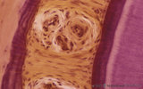 |
Cross section of tooth in alveolar bone - cat | Stain: Picric acid and hematoxylin. From left to right: alveolar bone covered with a woven type bone (bundle bone) tissue (stained darkly purple); yellow stained periodontal ligament with heavy collagen fibers (horizontal fibers) and blood vessels; bone-related part with Sharpey's fibers followed by... | oral cavity; alveolar bone | Poja Histology Collection - Oral Cavity Subset |
| 86 |
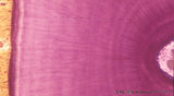 |
Cross-section of tooth in alveolar bone (cat, adult) | Stain: Picric acid and hematoxylin. From left to right: Periodontal ligament with blood vessels. Acellular cementum (dark purple rim). Dentin with radiair arrangement of dentinal tubules; fine incremental (imbrication) lines of von Ebner run at right angles to these tubules. These lines represent di... | oral cavity; incremental lines; von Ebner | Poja Histology Collection - Oral Cavity Subset |
| 87 |
 |
Cystic corpuscle in thymus (human, adult) | Stain: Azan. With age the remaining Hassall's corpuscles become more prominent and might represent large multiform structures of over hundreds of microns. The central cells (*) are keratinized. They might be swollen, calcify and become necrotic eventually, or undergo lysis, leaving a large cystic st... | thymic corpuscle (Hassalls); epithelioreticular cell (ERC); lymphoid tissue | Poja Histology Collection - Lymphatic Tissues and Organs Subset |
| 88 |
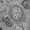 |
Cystic corpuscle in thymus (mouse, young adult) | Electron microscopy. This cystic corpuscle is lined by specialized epithelioreticular cells and exhibits a lumen (1) which is filled with long microvilli however, curiously derived from a flattened epithelial cell (2, nucleus). The cell contains few electron-dense keratohyalin granules (3). (-->) po... | epithelioreticular cells; thymic corpuscle; thymic cyst; lymphoid tissue | Poja Histology Collection - Lymphatic Tissues and Organs Subset |
| 89 |
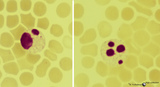 |
Degenerating granulocytes in peripheral blood smear (human) | Stain: May-Grnwald-Giemsa (MGG).Two neutrophils with degenerated nuclei. Note the pyknotic pattern (like mature erythroblast) of the separated and rounded nuclear lobes and the granules in the cytoplasm. Normally only a small number of granulocytes die in the blood. | Poja Histology Collection - Blood & Bone Marrow Subset | |
| 90 |
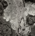 |
Dendritic cells in spleen (mouse) | Electron microscopy. The interdigitations (*) of the left dendritic cells (antigen-presenting cell or APC) (1 and 2a) are clearly shown. The right (2b) is a neighbour dendritic cell sectioned at the level of the Golgi area. (3) lymphocyte and (4) reticular cell. | electron microscopy; dendritic cell | Poja Histology Collection - Lymphatic Tissues and Organs Subset |
| 91 |
 |
Dendritic cells in spleen (rat) | Electron microscopy. The interdigitating dendritic cells (1) (so-called antigen-presenting cell, APC) exhibit numerous slender cell projections (1). (2) shows a macrophage with large lysosomes with heterogeneous contents. Small elongated fibroblastic reticular cells (3) form a structural framework ... | electron microscopy; dendritic cell | Poja Histology Collection - Lymphatic Tissues and Organs Subset |
| 92 |
 |
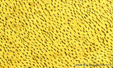
Dentin in cross section of tooth (human, adult) | Stain: Hematoxylin and eosin. Longitudinally sectioned dentinal tubules are parallely arranged, and numerous side branches are visible. | oral cavity; dentinal tubules | Poja Histology Collection - Oral Cavity Subset |
| 93 |
 |
Dentin in cross section of tooth - human, adult | Stain: Hematoxylin and eosin. Cross-sectioned dentinal tubules demonstrate: a light stained center with the odontoblastic process (Tomes' fiber); darker stained peritubular dentin (highly mineralized), also called Neuman's sheath. Intertubular dentin (less mineralized) is present between the tubule... | oral cavity; dentinal tubules; secondary dentin | Poja Histology Collection - Oral Cavity Subset |
| 94 |
 |
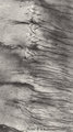
Dentinal tubules in longitudinal section of tooth - human, adult. Thin ground section, polarizing microscopy - optical axes of the polarizing plates are crossed at 60. | Slightly oblique section shows dentinal tubules depicted by brown-stained hollow fiber-like structures containing odontoblastic processes (Tomes' fibers) in a semi-three dimensional way. Numerous fine secondary branches of these processes are anastomosing with those of neighboring tubules. | oral cavity; Tomes' Fibers; dentinal tubules | Poja Histology Collection - Oral Cavity Subset |
| 95 |
 |
Dentinal tubules in longitudinal section of tooth - human, adult. Thin ground section, polarizing microscopy - optical axes of the polarizing plates are crossed at 60. | Slightly oblique section shows dentinal tubules depicted by brown-stained hollow fiber-like containing odontoblastic processes (Tomes' fibers) in a semi-three dimensional way ('stubble-field' aspect). Darker stained clusters represent anastomosing secondary branches. | oral cavity; Tomes' Fibers; dentinal tubules | Poja Histology Collection - Oral Cavity Subset |
| 96 |
 |
Dentinal tubules in longitudinal section of tooth - human, adult. Thin ground section, polarizing microscopy - optical axes of the polarizing plates are crossed at 60. | Slightly oblique section shows dentinal tubules depicted by brown-stained stubble structures containing odontoblastic processes (Tomes' fibers) in a semi-three dimensional way. Numerous fine branches of these processes are obvious in dentin. | oral cavity; Tomes' Fibers; dentinal tubules | Poja Histology Collection - Oral Cavity Subset |
| 97 |
 |
Dentinoenamel junction in longitudinal section of tooth (human, adult). Thin ground section. | From left to right: Enamel with fine striation (course of enamel rods or prisms). Few enamel tufts (left, dark) consisting of hypocalcified enamel rods and interprismatic substance arise from the junction. Scallop-like course of dentinoenamel junction. Dentin with dentinal tubules to the dentinoenam... | oral cavity; enamel tufts; dentinoenamel junction; dentinal tubules | Poja Histology Collection - Oral Cavity Subset |
| 98 |
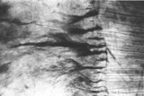 |
Dentinoenamel junction in longitudinal section of tooth - human, adult. Thin ground section. | From left to right: enamel with fine striation (composed of stalks of enamel rods or prisms); enamel tufts (dark) arise at the dentinoenamel junction, and these tufts consist of hypocalcified enamel prisms and interprismatic substance; dentin with dentinal tubules up to the dentinoenamel. | oral cavity; enamel tufts; dentinoenamel junction; dentinal tubules | Poja Histology Collection - Oral Cavity Subset |
| 99 |
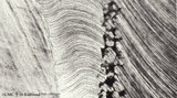 |
Dentinoenamel junction in the tooth (human, adult). Thin ground section of crown. | From left to right: Enamel with fine striation (composition of enamel rods or prisms); darker zones almost perpendicular to the striation are the incremental lines (Retzius) due to successive apposition of layers of enamel as the crown is formed. Dentinoenamel junction is shown as a narrow fissure f... | oral cavity; dentinal tubules; dentinoenamel junction; interglobular dentin; lines of Retzius | Poja Histology Collection - Oral Cavity Subset |
| 100 |
 |
Dentinoenamel junction in the tooth - human, adult. Thin ground section of crown. | From left to right: Superficial dentin (bluish) in the crown with S-shaped course of dentinal tubules; They pass uninterrupted through the irregular black structures (due to filling with air) representing hypocalcified areas (interglobular dentin); Mineralization of dentin starts in small globular ... | oral cavity; dentinal tubules; dentinoenamel junction; interglobular dentin; lines of Retzius | Poja Histology Collection - Oral Cavity Subset |