AAO-NANOS Neuro-Ophthalmology Clinical Collection: Derived from the AAO-NANOS Clinical Neuro-Ophthalmology collection produced on CD. The images are of selected cases from the NANOS teaching slide exchange, and the CD was produced under the direction of Larry Frohman, MD and Andrew Lee, MD.
The American Academy of Ophthalmology (AAO); The North American Neuro-Ophthalmology Association (NANOS).
NOVEL: https://novel.utah.edu/
TO
| Title | Creator | Description | ||
|---|---|---|---|---|
| 76 |
 |
Systemic Disorders With Optic Nerve and Retinal Findings | Joel M. Weinstein, MD | This 46-year-old woman noted progressive bilateral visual loss over a 10-month period. She had a malignant melanoma removed from her right thigh 2 years ago and excisional biopsy of an inguinal node metastasis 8 months ago. She also complained of poor night vision and rare intermittent ""sparkles"" ... |
| 77 |
 |
Systemic Disorders With Optic Nerve and Retinal Findings | Joel M. Weinstein, MD | This 46-year-old woman noted progressive bilateral visual loss over a 10-month period. She had a malignant melanoma removed from her right thigh 2 years ago and excisional biopsy of an inguinal node metastasis 8 months ago. She also complained of poor night vision and rare intermittent ""sparkles"" ... |
| 78 |
 |
Systemic Disorders With Optic Nerve and Retinal Findings | Joel M. Weinstein, MD | This 46-year-old woman noted progressive bilateral visual loss over a 10-month period. She had a malignant melanoma removed from her right thigh 2 years ago and excisional biopsy of an inguinal node metastasis 8 months ago. She also complained of poor night vision and rare intermittent ""sparkles"" ... |
| 79 |
 |
Neuro-Ophthalmic Consequences of Therapy | Joel M. Weinstein, MD | The patient is a 38-year-old woman with oligodendroglioma of the left cerebral hemisphere. The patient received intracarotid BCNU and developed BCNU retinopathy, optic neuropathy, and partial ophthalmoplegia. Image 92_22 Shows late changes, with more severe retinal ischemia, vascular attenuation, an... |
| 80 |
 |
Systemic Disorders With Optic Nerve and Retinal Findings | Larry P. Frohman, MD | A 29-year-old African American woman presented with headaches, bilateral transient visual obscurations, blurred vision, numbness, and weakness of the lower extremities with myalgia and joint pains. She had an unplanned 12-pound weight loss over 2 months. A neurologist and internist diagnosed her wit... |
| 81 |
 |
Motility Disturbances | Larry P. Frohman, MD | This 5-year-old child presented with a 70 PD exotropia OS and a right face turn. She had a CT scan of the head at age 4 months that was normal , and she was felt to have an isolated left medial rectus paresis. Her acuity was 20/20 OU. She could fuse with a large face turn, and was orthomorphic is ex... |
| 82 |
 |
Motility Disturbances | Larry P. Frohman, MD | This 5-year-old child presented with a 70 PD exotropia OS and a right face turn. She had a CT scan of the head at age 4 months that was normal , and she was felt to have an isolated left medial rectus paresis. Her acuity was 20/20 OU. She could fuse with a large face turn, and was orthomorphic is ex... |
| 83 |
 |
Motility Disturbances | Larry P. Frohman, MD | This 5-year-old child presented with a 70 PD exotropia OS and a right face turn. She had a CT scan of the head at age 4 months that was normal , and she was felt to have an isolated left medial rectus paresis. Her acuity was 20/20 OU. She could fuse with a large face turn, and was orthomorphic is ex... |
| 84 |
 |
Motility Disturbances | Larry P. Frohman, MD | This 5-year-old child presented with a 70 PD exotropia OS and a right face turn. She had a CT scan of the head at age 4 months that was normal , and she was felt to have an isolated left medial rectus paresis. Her acuity was 20/20 OU. She could fuse with a large face turn, and was orthomorphic is ex... |
| 85 |
 |
Systemic Disorders With Optic Nerve and Retinal Findings | Larry P. Frohman, MD | A 29-year-old African American woman presented with headaches, bilateral transient visual obscurations, blurred vision, numbness, and weakness of the lower extremities with myalgia and joint pains. She had an unplanned 12-pound weight loss over 2 months. A neurologist and internist diagnosed her wit... |
| 86 |
 |
Systemic Disorders With Optic Nerve and Retinal Findings | Larry P. Frohman, MD | A 35-year-old African-American woman had gradual bilateral painless visual loss over 3 months. When initially seen, the visual acuities were HM OD, NLP OS. The MRI showed diffused enhancement of the optic nerves and lacrimal glands. The evaluation strongly suggested sarcoidosis, with elevated angiot... |
| 87 |
 |
Systemic Disorders With Optic Nerve and Retinal Findings | Larry P. Frohman, MD | A 25-year-old African-American woman presented with a 20/200 optic neuropathology and no other illness. Because this disc appearance of a granuloma led us to suspect occult sarcoidosis, she underwent systemic evaluation. She was ultimately shown to have systemic sarcoidosis, including pulmonary invo... |
| 88 |
 |
Isolated Optic Neuritis/Neuropathy | Anthony C. Arnold, MD | This 48-year-old man presented with a 1-month history of headache. Both discs had the appearance seen in this image, with prominent peripapillary nerve fiber layer myelination; the disc itself is hyperemic, with dilated, telangiectatic surface vasculature, suggesting true disc edema as well. |
| 89 |
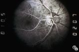 |
Optic Disc Drusen, Fluorescein Angiogram | Anthony C. Arnold, MD | Images 92_64 and 92_67 demonstrate the characteristics of optic disc drusen on flourescein angiography. This image shows the early arteriovenous phase, with irregular dye uptake and focal hypoflourescence superotemporally. Pair with 92_63 and 92_67 |
| 90 |
 |
Ocular Manifestations of Systemic Disorders | Rosa A. Tang, MD | Myasthenia gravis should be considered in any patient with painless, pupil-spared, nonapoptotic ophthalmoplegia. It may mimic any ophthalmoparesis. Involvement of the medical rectus may result in a pseudointernuclear ophthalmoplegia. Pair with 96_24 and 96_25. |
| 91 |
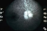 |
Optic Nerve Drusen, Late Fluorescein Angiogram | Anthony C. Arnold, MD | Images 92_64 and 92_67 demonstrate the characteristics of optic disc drusen on flourescein angiography. This image displays the nodular staining of the drusen without leakage. Pair with 92_63 and 92_64. |
| 92 |
 |
Motility Disturbances | Don Bienfang, MD | This patient displays a posttraumatic left fourth nerve palsy sustained after having struck her head on the dashboard. |
| 93 |
 |
Motility Disturbances | Don Bienfang, MD | This patient displays a posttraumatic left fourth nerve palsy sustained after having struck her head on the dashboard. |
| 94 |
 |
Motility Disturbances | Don Bienfang, MD | This patient displays a posttraumatic left fourth nerve palsy sustained after having struck her head on the dashboard. |
| 95 |
 |
Systemic Disorders With Optic Nerve and Retinal Findings | Larry P. Frohman, MD | A 35-year-old African-American woman had gradual bilateral painless visual loss over 3 months. When initially seen, the visual acuities were HM OD, NLP OS. The MRI showed diffused enhancement of the optic nerves and lacrimal glands. The evaluation strongly suggested sarcoidosis, with elevated angiot... |
| 96 |
 |
Ocular Manifestations of Systemic Disorders | Larry P. Frohman, MD | A 17-year-old girl had undergone multiple superficial biopsies of the orbit for what was felt to be refractory orbital pseudotumor. Initial evaluation revealed the saddle-nose deformity, which the patient confirmed was acquired. More extensive biopsy was consistent with lymphomatoid granulomatosis. ... |
| 97 |
 |
Ocular Manifestations of Congenital/Inherited Diseases | Jacqueline A. Leavitt, MD | This 22-year-old woman has neurofibromatosis, type 2. Acuity, color plates, pupillary responses, slit-lamp examination, IOP, fields, and funduscopy are all normal. There is a 3 mm proptosis OS. The patient has recently undergone gamma knife for the acoustic tumor, and she has residual facial nerve p... |
| 98 |
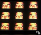 |
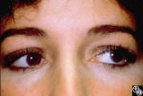
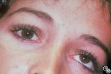
Motility Disturbances | Rosa A. Tang, MD | Traumatic damage to the third cranial nerve may result in aberrant regeneration of fibers that innervate the eyelid, pupil, or extraocular muscles. For instance, there may be lid retraction in attempted downgaze. Any combination of aberrant activation of third nerve-innervated structures may occur, ... |
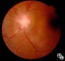
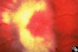
| 99 |
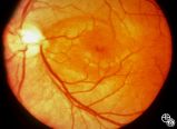 |
Isolated Congenital Optic Disc Anomalies | Thomas R. Wolf, MD | An optic pit is a small defect in the optic disc that may be asymptomatic in isolation. Patients may develop an associated serous detachment of the macula. The condition is usually unilateral but may be bilateral. A fluorescein angiogram may demonstrate the serous detachment, and laser photocoagulat... |
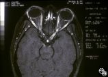
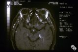
| 100 |
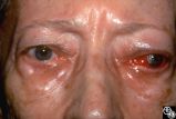 |
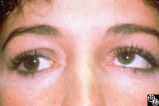
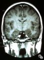
Ocular Manifestations of Systemic Disorders | Rosa A. Tang, MD | Thyroid eye disease may cause proptosis and extraocular muscle enlargement that may be seen on orbital imaging studies. In general, coronal images allow the best visualization of the extraocular muscle enlargement. Pair with 94_45 and 94_46. |