The Health Education Assets Library (HEAL) is a collection of over 22,000 freely available digital materials for health sciences education. The collection is now housed at the University of Utah J. Willard Marriott Digital Library.
Filters: Collection: ehsl_heal
| Title | Description | Subject | Collection | ||
|---|---|---|---|---|---|
| 51 |
 |
Exocrine gland - striated (intralobular) duct - salivary glands, pancreas | Scheme electronmicroscopy. Part of a striated duct with basally membrane infoldings and mitochondria accumulations in the tall columnar cells. Small junctional complexes as well as apically mitochondria and numerous vesicles are present. | oral cavity; striated duct | Poja Histology Collection - Oral Cavity Subset |
| 52 |
 |
Exocrine gland - striated (intralobular) ducts - parotid gland, human | Stain: Azan. Cross-sections of striated ducts in upper part between serous acini. Note at the right bottom part of (lighter stained) lining cells of an intercalated duct. Connective tissues between the structures are blue stained. | oral cavity; striated duct | Poja Histology Collection - Oral Cavity Subset |
| 53 |
 |
Exocrine gland - submandibular gland, gerbil | Electronmicroscopy. Part of a serous acinus of this mixed gland. Note characteristic electron-dense secretion granules, the mature ones are larger. At the lower bottom is shown part of a lining cell of the intercalated duct. | oral cavity; serous gland | Poja Histology Collection - Oral Cavity Subset |
| 54 |
 |
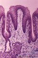
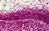
Filiform papillae of the tongue (dorsal side, human) | Stain: Heidenhain light bordeaux. Detail of the threadlike keratinized extensions of the stratified epithelium. Primary connective tissue papillae with 2 to 3 secondary papillae. Note the absence of taste buds in filiform papillae. | oral cavity; filiform papillae | Poja Histology Collection - Oral Cavity Subset |
| 55 |
 |
Filiform papillae of the tongue (dorsal side, human) | Stain: Azan. Oblique cross-section through the top of the filiform papillae. At the top of the picture keratinized extensions. Primary connective tissue papillae (blue) divide in several small secondary ones. | oral cavity; filiform papillae | Poja Histology Collection - Oral Cavity Subset |
| 56 |
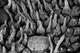 |
Filiform papillae of the tongue (dorsal side, human, neonate) | Scanning electronmicroscopy. Slender, thin threadlike extensions of the filiform papillae. In between a broad fungiform papilla. | oral cavity; filiform papillae; fungiform papilla | Poja Histology Collection - Oral Cavity Subset |
| 57 |
 |
Filiform papillae of the tongue - dorsal side, human | Stain: Goldner trichrome. Detail of the threadlike keratinized extensions (red) of the stratified epithelium. Primary connective tissue papillae (green) divide in several small secondary ones. Note the absence of taste buds in filiform papillae. | oral cavity; filiform papillae | Poja Histology Collection - Oral Cavity Subset |
| 58 |
 |
Filiform papillae of the tongue - dorsal side, human | Stain: Heidenhain light bordeaux. Transverse section through the middle of the filiform papillae showing secondary connective tissue papillae (lightly stained). | oral cavity; filiform papillae | Poja Histology Collection - Oral Cavity Subset |
| 59 |
 |
Foliate papillae of the tongue (dorsal side, rabbit) | Stain: Goldner trichrome. Foliate papil with primary and secondary connective tissue projections. Taste buds are localized in the lining, non-keratinized epithelium of the grooves. The serous gland of von Ebner (minor salivary gland) drains via an enlarged duct into the left groove of the papil. | oral cavity; foliate papillae; von Ebner | Poja Histology Collection - Oral Cavity Subset |
| 60 |
 |
Free denticulus (pulp stone) in the tooth - longitudinal section of root pulp; human, adult | Stain: Hematoxylin and eosin. In the center of pulp connective tissue a free pulp stone (false denticulus). Dispersed through the pulp several small stones. Left of the central stone a longitudinal sectioned thin-walled blood vessel; right of the stone bundles of nerve fibers. | oral cavity; denticulus; pulp stone | Poja Histology Collection - Oral Cavity Subset |
| 61 |
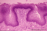 |
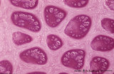
Fungiform papillae of the tongue (dorsal side, human) | Stain: Hematoxylin and eosin. A broad prominent papil of connective tissue (without secondary papils) covered by non-keratinized stratified epithelium. This specimen does not show any taste bud. Well vascularized lamina propria. | oral cavity; fungiform papillae | Poja Histology Collection - Oral Cavity Subset |
| 62 |
 |
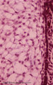
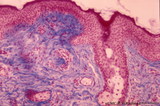
Gingiva ('attached' gingiva of decalcified alveolar bone, human, adult) | Stain: Hematoxylin and eosin. Thick layer of stratified squamous epithelium with parakeratosis (reddish) and shallow papillae of the lamina propria. At the bottom lamellar bone tissue (alveolar bone) with thickened periosteum. | oral cavity; alveolar bone | Poja Histology Collection - Oral Cavity Subset |
| 63 |
 |
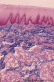
Gingiva ('free' gingiva of decalcified tooth, human, adult) | Stain: Hematoxylin and eosin. Thick layer of stratified squamous epithelium with parakeratosis (reddish) and many narrow papillae of the lamina propria. | oral cavity | Poja Histology Collection - Oral Cavity Subset |
| 64 |
 |
Gingiva - 'attached' gingiva of decalcified alveolar bone, human | Stain: Hematoxylin and eosin. Stratified squamous epithelium with parakeratosis (reddish) and deep papillae of the lamina propria. At the bottom, lamellar bone tissue (alveolar bone) with thickened periosteum (part of the periodontal ligament). At the bottom dentin. | oral cavity | Poja Histology Collection - Oral Cavity Subset |
| 65 |
 |
Gingiva - 'free' gingiva of decalcified alveolar bone, human | Stain: Hematoxylin and eosin. Thick layer of stratified squamous epithelium with parakeratosis (reddish) and broad papillae of the lamina propria. At the left, dense collagen tissue (projections of the the periodontal ligament). | oral cavity | Poja Histology Collection - Oral Cavity Subset |
| 66 |
 |
Late cap stage in tooth development - human, embryo | Stain: Azan. From top to bottom: Top side stellate reticulum (enamel pulp) consisting of a network of ectoderm-derived cells; Right side outer dental epithelium with part of the fibrous tooth follicle. This epithelium will further develop downwards as the outer layer of the Hertwig's epithelial root... | oral cavity | Poja Histology Collection - Oral Cavity Subset |
| 67 |
 |
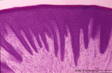
Late cap stage in tooth development - human, embryo | Stain: Azan. The stellate reticulum (enamel pulp) consists of a network of ectoderm-derived branched cells and fluid-filled spaces (a.o. proteoglycans). It is a specialized avascular layer as a support and protection for the inner dental epithelial cells. At the right side one layer of cuboidal oute... | oral cavity; tooth development; outer dental epithelium; stellate reticulum | Poja Histology Collection - Oral Cavity Subset |
| 68 |
 |
Late cap stage in tooth development - human, embryo | Stain: Azan. From top to bottom: At the top stellate reticulum (enamel pulp) consisting of a loose network of ectoderm-derived cells; Darker stained cell layers of the stratum intermedium; Columnar inner dental epithelium (presecreetory ameloblasts) at the distal side (secretion area) oriented towar... | oral cavity | Poja Histology Collection - Oral Cavity Subset |
| 69 |
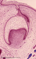 |
Late cap stage of tooth development - human, embryo; low magnification | Stain: Azan. From top to bottom: Stratified ectoderm with a distinct basal layer (red line) of cuboid cells; Dental lamina giving rise to the cap stage (center) and to the primordium of permanent tooth (right); Odontogenic organ or enamel organ (future deciduous tooth surrounded by fibrous tooth fol... | oral cavity; dental lamina | Poja Histology Collection - Oral Cavity Subset |
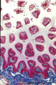
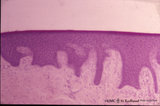
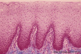
| 70 |
 |
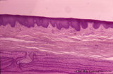
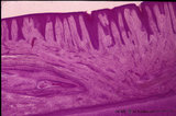
Lip (human), external skin surface | Stain: Azan. Keratinized squamous epithelium with one hair follicle associated with sebaceous gland. Richly vascularized connective tissue. Note the thin red cornified layer on the surface. | oral cavity | Poja Histology Collection - Oral Cavity Subset |
| 71 |
 |
Lip (human), mucoserous labial glands in mucous inner surface | Stain: Azan. Mixed labial glands with serous demilunes (von Ebner-Giannuzzi), a few myoepithelial cells, and a striated duct (right upper corner). | oral cavity; lining mucosa; labial glands | Poja Histology Collection - Oral Cavity Subset |
| 72 |
 |
Lip (human), mucous inner surface | Stain: Azan. Non-keratinized epithelium with high, narrow dermal papillae and many capillaries. Mixed labial glands (seromucous) and few skeletal muscle fibers (orbicularis oris) in the submucosa. | oral cavity; lining mucosa; labial glands | Poja Histology Collection - Oral Cavity Subset |
| 73 |
 |
Lip (human), mucous inner surface | Stain: Azan. Non-keratinized epithelium with high, narrow dermal papillae and many capillaries. Note lymphocytic infiltrates and the capillaries in the dermal papillae. | oral cavity; lining mucosa | Poja Histology Collection - Oral Cavity Subset |
| 74 |
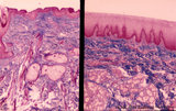 |
Lip (human), outer (left) and inner side (right) | Stain: Azan. Left image: keratinized squamous epithelium with thin red cornified layer, hair follicle, sebaceous glands and skeletal muscle fibers (orbicularis oris). Right image: non-keratinized epithelium with high, narrow dermal papillae and more capillaries. Mixed labial glands (seromucous), f... | oral cavity; lining mucosa | Poja Histology Collection - Oral Cavity Subset |
| 75 |
 |
Lip (human), region between red zone (vermilion border) and mucosa inner surface | Stain: Azan. Bundles of skeletal muscle fibers (musculus orbicularis oris), highly vascularized lamina propria. Note the narrow dermal papillae on the left side (inner lip) and the broad irregular papillae on the right side (red zone of the lip). | oral cavity; lining mucosa; red zone; vermilion border | Poja Histology Collection - Oral Cavity Subset |