Best known for his world-renowned neuro-ophthalmology unit based at the University of California, San Francisco, William Hoyt, MD collected here more than 850 of his best images covering a wide range of disorders.
William F. Hoyt, MD, Professor Emeritus of Ophthalmology, Neurology and Neurosurgery, Department of Ophthalmology, University of California, San Francisco.
NOVEL: https://novel.utah.edu/
TO
Filters: Collection: "ehsl_novel_wfh"
| Title | Description | Type | ||
|---|---|---|---|---|
| 51 |
 |
Retinal Signs of Atheromatous Embolization | Retinal signs of atheromatous embolization. Series shows event in progress. R3_A19d shows progress along the same transit path for both emboli. Anatomy: Retina. Pathology: Carotid atheromatous disease. Disease/Diagnosis: Carotid atheromatous vascular disease. Clinical: No visual symptom. | Image |
| 52 |
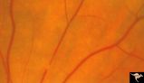 |
Retinal Signs of Atheromatous Embolization | Retinal signs of atheromatous embolization. Series shows event in progress. R3_A19a shows atheromatous embolus traveling up the arteriole. Anatomy: Retina. Pathology: Carotid atheromatous disease. Disease/Diagnosis: Carotid atheromatous vascular disease. Clinical: No visual symptom. | Image |
| 53 |
 |
Retinal Signs of Atheromatous Embolization | Retinal signs of atheromatous embolization. Series shows event in progress. R3_A19b shows embolus approaching the bifurcation. Anatomy: Retina. Pathology: Carotid atheromatous disease. Disease/Diagnosis: Carotid atheromatous vascular disease. Clinical: No visual symptom. | Image |
| 54 |
 |
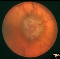
A201 Disc Swelling with Big Blind Spot Syndrome | Blind spot larger than could be explained by visible edema. Subretinal white dots probably indicate margin of blind spot. Anatomy: Optic disc; Retina. Pathology: Unknown. Disease/ Diagnosis: Big blind spot syndrome. Clinical: symptoms: photosias, blurred vision signs: Disc swelling; white spots in t... | Image |
| 55 |
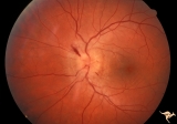 |
A202 Disc Swelling with Big Blind Spot Syndrome | Blind spot larger than could be explained by visible disc edema. Reference: Fletcher WA, Imes RK, Goodman D, Hoyt WF. Acute idiopathic blind spot enlargement. A big blind spot disc edema. Arch Ophthalmol. 1988 Jan;106(1):44-9. Anatomy: Optic disc; Retina. Pathology: Unknown. Disease/ Diagnosis: Big ... | Image |
| 56 |
 |
A203 Disc Swelling with Big Blind Spot Syndrome | Slight inferior swelling in patient with grossly enlarged blind spot. 66 year old woman. Anatomy: Optic disc; Retina. Pathology: Unknown. Disease/ Diagnosis: Big blind spot syndrome. Clinical: symptoms: photopsias; blurred vision signs: disc swelling; white dots in the retina; enlarged blind spot on... | Image |
| 57 |
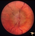 |
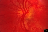
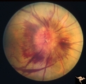
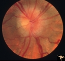
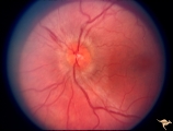
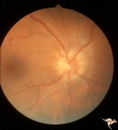
Bilateral Chronic Papilledema | Left eye. Frisen's stage 5. Patient with long standing aqueductal stenosis. Bilateral Chronic Papilledema. Man. Anatomy: Optic disc. Pathology: Papilledema. Disease/Diagnosis: Papilledema from aqueductal stenosis. | Image |
| 58 |
 |
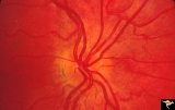
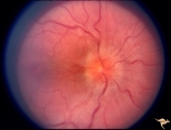
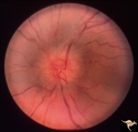
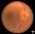
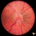
Bilateral Chronic Papilledema | Right eye. Frisen's stage 5. Patient with long standing aqueductal stenosis. Bilateral Chronic Papilledema. Man. Anatomy: Optic disc. Pathology: Papilledema. Disease/Diagnosis: Papilledema from aqueductal stenosis. | Image |
| 59 |
 |
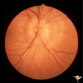
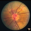
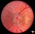
Bilateral Crowded Discs | Left eye. Bilateral crowded discs with congenital blurring. Blurred disc margins are not from edema. Note optic cup is absent. Pair with right eye in PP_1a, and brother in PP_2. Mother has drusen of the optic disc in PP_11aa & b. Sister has drusen in PP_11c. Anatomy: Optic disc. Pathology: Normal va... | Image |
| 60 |
 |
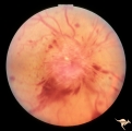
Bilateral Crowded Discs (Family) | Right eye. Bilateral crowded discs with congenital blurring. Blurred disc margins are not from edema. Note optic cup is absent. Pair with left eye in PP_1b, and brother in PP_2. Mother has drusen of the optic disc in PP_11a & b. Sister has drusen in PP_11c. Anatomy: Optic disc. Pathology: Normal var... | Image |
| 61 |
 |
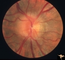
Bilateral Hemorrhagic Papilledema | Left eye. Bilateral Hemorrhagic Papilledema from cardio-respiratory disease. Woman. Anatomy: Optic disc. Pathology: Bilateral papilledema, hemorrhagic. Disease/Diagnosis: Pseudotumor due to cardio-respiratory disease. Clinical notes: Woman with headache, shortness of breath. | Image |
| 62 |
 |
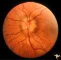
Bilateral Hemorrhagic Papilledema | Bilateral Hemorrhagic Papilledema from cardio-respiratory disease. Woman. Anatomy: Optic disc. Pathology: Bilateral papilledema, hemorrhagic. Disease/Diagnosis: Pseudotumor due to cardio-respiratory disease. Clinical notes: Woman with headache, shortness of breath. | Image |
| 63 |
 |
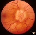
Bilateral Papilledema | Left eye. Has intra-retinal exudate and unusual vascular changes in the optic disc. Pre-pubertal girl. Anatomy: Optic disc. Pathology: Bilateral papilledema; exudative deposits in macula. Disease/Diagnosis: Pseudotumor. Clinical: Pubertal girl; headaches. | Image |
| 64 |
 |
Bilateral Papilledema | Right eye. Bilateral Papilledema in 410 pound man with tracheostomy for pulmonary insufficiency. Anatomy: Optic disc. Pathology: Papilledema. Disease/Diagnosis: Pseudotumor due to: sleep apnea due to cardiopulmonary insufficiency syndrome. Pickwickian syndrome. Clinical notes: Headache; obesity. | Image |
| 65 |
 |
Bilateral Papilledema | Right eye. Bilateral Papilledema in patient with cardiopulmonary insufficiency. Woman. Anatomy: Optic disc. Pathology: Papilledema. Disease/Diagnosis: cardiopulmonary insufficiency causing intracranial hypertension. Clinical notes: headache. | Image |
| 66 |
 |
Bilateral Papilledema | Right eye. Bilateral Papilledema in a patient with hyperthyroidism. Woman. Anatomy: Optic disc. Pathology: Papilledema. Disease/Diagnosis: Bilateral papilledema with hyperthyroidism. | Image |
| 67 |
 |
Bilateral Papilledema | Left eye. Bilateral Papilledema with hypoparathyroidism. Woman. Anatomy: Optic disc. Pathology: Papilledema. Papilledema with hypopararthyroidism. | Image |
| 68 |
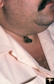 |
Bilateral Papilledema | Picture of patient. 410 pound man with tracheostomy done for sleep apnea due to cardiopulmonary insufficiency syndrome. Pickwickian syndrome. Anatomy: Optic disc. Pathology: Papilledema. Disease/Diagnosis: Pseudotumor due to: sleep apnea due to cardiopulmonary insufficiency syndrome. Pickwickian syn... | Image |
| 69 |
 |
Bilateral Papilledema | Right eye. Bilateral Papilledema from vitamin A toxicity. Vitamin A pseudotumor cerebri syndrome in a 25 year old weight lifter. Anatomy: Optic disc. Pathology: Bilateral papilledema. Disease/Diagnosis: Pseudotumor due to vitamin A toxicity and weight lifting. Clinical notes: Headache, weight lifter... | Image |
| 70 |
 |
Bilateral Papilledema | Left eye. Bilateral Papilledema in patient with cardiopulmonary insufficiency. Woman. Anatomy: Optic disc. Pathology: Papilledema. Disease/Diagnosis: Cardiopulmonary insufficiency causing intracranial hypertension. Clinical notes: Headache. | Image |
| 71 |
 |
Bilateral Papilledema | Right eye. Bilateral Papilledema with hypoparathyroidism. Woman. Anatomy: Optic disc. Pathology: Papilledema. Disease/Diagnosis: Papilledema with hypoparathyroidism. | Image |
| 72 |
 |
Bilateral Papilledema | Left eye. Bilateral Papilledema in a patient with hyperthyroidism. Woman. Anatomy: Optic disc. Pathology: Papilledema. Disease/Diagnosis: Bilateral papilledema. | Image |
| 73 |
 |
Bilateral Papilledema | Left eye. Bilateral Papilledema from vitamin A toxicity. Vitamin A pseudotumor cerebri syndrome in a 25 year old weight lifter. Anatomy: Optic disc. Pathology: Bilateral papilledema. Disease/Diagnosis: Pseudotumor due to vitamin A toxicity and weight lifting. Clinical notes: Headache, weight lifter. | Image |
| 74 |
 |
Bilateral Papilledema | Left eye. Chronic Bilateral Papilledema. Anatomy: Optic disc. Pathology: Chronic bilateral papilledema. Disease/Diagnosis: Pseudotumor long standing. Clinical notes: Chronic headache; Obesity. | Image |
| 75 |
 |
Bilateral Papilledema | Right eye. Pre-pubertal girl. Anatomy: Optic disc. Pathology: Bilateral papilledema; exudative deposits in macula. Disease/Diagnosis: Pseudotumor. Clinical notes: Pubertal girl; headaches. | Image |