Collection of materials relating to neuro-ophthalmology as part of the Neuro-Ophthalmology Virtual Education Library.
NOVEL: https://novel.utah.edu/
TO
- NOVEL720
| Title | Creator | Description | Subject | ||
|---|---|---|---|---|---|
| 51 |
 |
A Macro Dilemma: Demonstrations of the Anterior Chiasmal Syndrome and Wilbrand's Knee | Ariel Axelbaum, MD; Nurhan Torun, MD | Presentation reviewing two cases that demonstrate several cases of anterior chiasmal syndromes and the variability in patient presentations with sellar masses. | Anterior Chiasmal Syndromes; Masses of the Sella |
| 52 |
 |
Neuro-ophthalmic Disorders in Pregnancy: With an Eye to Future Eye Health | Kathleen B. Digre, MD | Presentation covering conditions relevant to neuro-ophthalmology, including vascular disorders that affect vision, Pseudotumor Cerebri Syndrome, venous sinus thrombosis, idiopathic intracranial hypertension, and severe pre-eclampsia and eclampsia. | Pregnancy |
| 53 |
 |
Computed Tomography (CT): Principles, Technique, and Neuro-ophthalmic Applications | Alex Fraser, MD | Presentation covering Computed Tomography principles, adverse effects, comparison vs. MRI, and assorted examples of neuro-ophthalmic interest. | Computed Tomography (CT) |
| 54 |
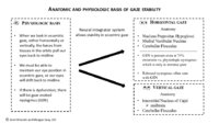 |
Anatomic and Physiologic Basis for Gaze Stability | Ariel Winnick and Meagan Seay, DO | Diagram describing the anatomic and physiologic basis of gaze stability. | Gaze Stability |
| 55 |
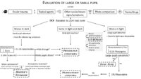 |
Pupil Evaluation Flowchart | Ariel Winnick; Eric Caskey, MD; Meagan Seay, DO | Flow chart outlining the evaluation of large or small pupils. | Pupil Evaluation |
| 56 |
 |
Heavy Eye Syndrome | Meagan D. Seay, DO; Bradley J. Katz, MD | A brief overview of heavy eye syndrome. | Heavy Eye Syndrome |
| 57 |
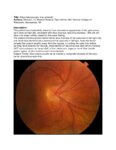 |
Situs Inversus Optic Disc Anomaly | Michael Hii, Medical Student; Ryan Walsh, MD | This patient was incidentally-noted to have anomalous appearance of the optic discs, right more so than left, consistent with situs inversus optic disc anomaly. She did not have any visual deficits related to this exam finding. ; The patient's fundus photos demonstrate situs inversus of the optic ... | Situs Inversus Optic Disc Anomaly |
| 58 |
 |
Superior Segmental Optic Disc Hypoplasia (SSOH) "Topless Disc Syndrome" | Sparsh Jain, Medical Student; Ryan Walsh, MD | This is a case of superior segmental optic disc hypoplasia that was found incidentally after a screening visual field test revealed an asymptomatic inferior field defect in the left eye. The patient has a unilateral SSOH in the left eye. | Superior Segmental Optic Disc Hypoplasia (SSOH) |
| 59 |
 |
Suprasellar Meningioma | Sumayya Almarzouqi, MD | Description of a case of suprasellar or sellar mass causeing chiasmal compression. | Suprasellar Meningioma |
| 60 |
 |
Brown Syndrome | Meagan Seay, DO | A brief overview of Brown Syndrome. | Brown Syndrome |
| 61 |
 |
Bergmeister Papilla | Sumayya Almarzouqi, MD | A brief overview of Bergmeister papilla, a rare congenital disc anomaly. It arises from the center of the optic disc consists of a small tuft of fibrous tissue and represents a remnant of the fetal hyaloid artery. | Bergmeister Papilla |
| 62 |
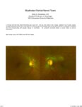 |
Myelinated Retinal Nerve Fibers | Scott N. Grossman, MD | A 33 year old man has noted chronically poor vision OS - left eye color noted to be 'orange' instead of red. fundus photos revealed myelinated retinal nerve fiber layer OU (OS>OD) with corresponding linear paracentral scotoma on Humphrey visual field 24-2 OS corresponding with greatest degree of my... | Myelinated Retinal Nerve Fibers |
| 63 |
 |
Modern Imaging of Optic Disc Drusen | Meagan Seay, DO | This is a short powerpoint describing imaging techniques (specifically OCT-EDI, fundus autofluorescence, and B-scan ultrasonography) for optic disc drusen. Examples of these techniques are included. | Optic Disc Drusen; Imaging; OCT-EDI; Fundus Autofluorescence; B-scan Ultrasonography |
| 64 |
 |
Congenital Hydrocephalus | Mays El-Dairi, MD | Presentation covering an overview of congenital hydrocephalus. | Congenital Hydrocephalus |
| 65 |
 |
Tolosa Hunt Syndrome | Sahil Aggarwal, MD; Jason Liss, MD | Presentation covering an overview of Tolosa Hunt Syndrome. | Tolosa Hunt Syndrome |
| 66 |
 |
Fibrous Dysplasia | Mays El-Dairi, MD | Presentation covering an overview of fibrous dysplasia. | Fibrous Dysplasia |
| 67 |
 |
Muscular Dystrophy Classification | Brian Villafuerte, MD, Ezequiel Piccione, MD | Presentation covering an overview of muscular dystrophy classification. | Muscular Dystrophy Classification |
| 68 |
 |
Myotonic Dystrophy | Brian Villafuerte, MD, Ezequiel Piccione, MD | Presentation covering an overview of myotonic dystrophy. | Myotonic Dystrophy |
| 69 |
 |
Night Wolf | Mehdi Tavakoli, MD; Byron Lam, MD | A case presentation on radiation optic neurology. | Radiation Optic Neuropathy |
| 70 |
 |
Cotton Wool Spots: The Basics | Arnav Gupta, BHSc; Rahul Sharma, MD, MPH | A presentation describing cotton wool spots, an abnormal finding on funduscopic exam of the retina of the eye. | Cotton Wool Spots; Retina |
| 71 |
 |
Disc Cupping: The Basics | Arnav Gupta, BHSc; Rahul Sharma, MD, MPH | A presentation describing optic disc cupping, due to damage of optic nerve fibres. | Disc Cupping; Optic Disc |
| 72 |
 |
Retinal Detachment: The Basics | Arnav Gupta, BHSc; Rahul Sharma, MD, MPH | A presentation describing retinal detachment. | Retinal Detachment |
| 73 |
 |
Retinal Exudate: The Basics | Arnav Gupta, BHSc; Rahul Sharma, MD, MPH | A presentation describing retinal exudate. | Retinal Exudate |
| 74 |
 |
Retinal Hemorrhage: The Basics | Arnav Gupta, BHSc; Rahul Sharma, MD, MPH | A presentation describing retinal hemorrhage. | Retinal Hemorrhage |
| 75 |
 |
Age-related Macular Degeneration: The Basics | Arnav Gupta, BHSc; Rahul Sharma, MD, MPH | A presentation covering age-related macular degeneration ("ARMD" or "AMD"), an acquired, progressive, chronic, degenerative disease of the retina. | Macular Degeneration |